

Spatial vulnerability: bacterial arrangements, microcolonies, and biofilms as responses to low rather than high phage densities
- PMID: 22754643
- PMCID: PMC3386622
- DOI: 10.3390/v4050663
Spatial vulnerability: bacterial arrangements, microcolonies, and biofilms as responses to low rather than high phage densities
Abstract
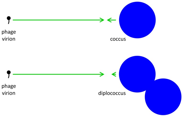
The ability of bacteria to survive and propagate can be dramatically reduced upon exposure to lytic bacteriophages. Study of this impact, from a bacterium's perspective, tends to focus on phage-bacterial interactions that are governed by mass action, such as can be observed within continuous flow or similarly planktonic ecosystems. Alternatively, bacterial molecular properties can be examined, such as specific phage‑resistance adaptations. In this study I address instead how limitations on bacterial movement, resulting in the formation of cellular arrangements, microcolonies, or biofilms, could increase the vulnerability of bacteria to phages. Principally: (1) Physically associated clonal groupings of bacteria can represent larger targets for phage adsorption than individual bacteria; and (2), due to a combination of proximity and similar phage susceptibility, individual bacteria should be especially vulnerable to phages infecting within the same clonal, bacterial grouping. Consistent with particle transport theory-the physics of movement within fluids-these considerations are suggestive that formation into arrangements, microcolonies, or biofilms could be either less profitable to bacteria when phage predation pressure is high or require more effective phage-resistance mechanisms than seen among bacteria not living within clonal clusters. I consider these ideas of bacterial 'spatial vulnerability' in part within a phage therapy context.
Keywords: adsorption; bacteriophage; biofilms; cellular arrangements; ecology; microcolonies; particle transport; phage therapy; phages.
Figures
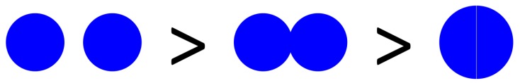
 . Twice its volume (V2) therefore is
  , which as a sphere is equal to
  . For
. For  , then r2 = 21/3r1. With such shading, then, diameter increases by only 21/3 = 1.26 fold.
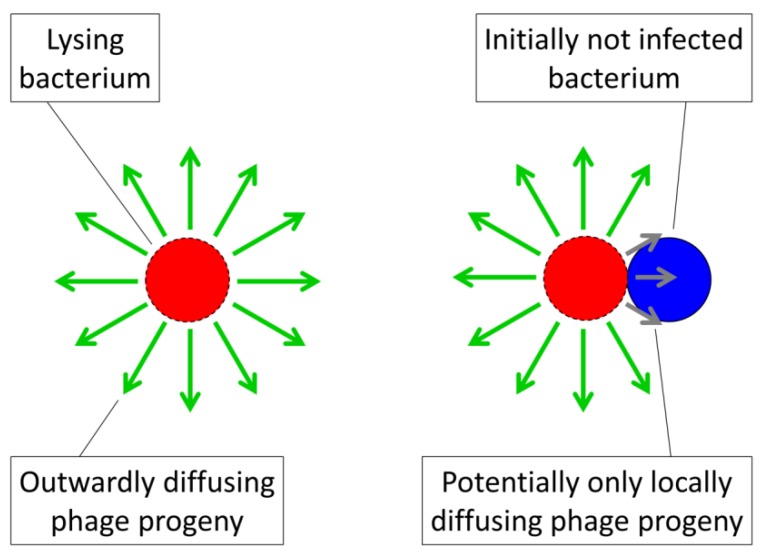
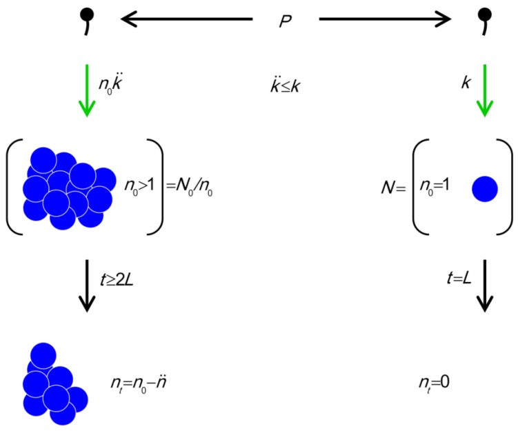
 (phage adsorption constant considering reductions due to shading of bacteria by bacteria found within bacterial arrangements), n (number of bacteria found per arrangement), N (bacterial density of overall environment), L (phage latent period, which is the duration of a phage infection), and
  (number of bacteria per arrangement lost subsequent to phage infection of one cell in the arrangement). Likelihood of phage adsorption of bacterial arrangements is n and density of arrangements within environments is equal to N/n = N0/n (or indeed n0 and N0/n0, respectively, to reflect that n changes as a function of time in the figure). The inequality t ≥ 2L indicates how phage acquisition of bacteria within a bacterial arrangement, according to this model, involves at least two sequential rounds of phage infection. The absence of cells in the lower right is intentional as too is the reduction in cell number to nt in the lower left. Both of these reductions in cell number, going from middle to bottom, indicate phage-induced bacterial lysis.
References
-
- Stoodley P., Sauer K., Davies D.G., Costerton J.W. Biofilms as complex differentiated communities. Ann. Rev. Microbiol. 2002;56:187–209. - PubMed
-
- Kjelleberg S., Givskov M. The Biofilm Mode of Life: Mechanisms and Adaptations. Horizon Biosciences; Norfolk, UK: 2007.
-
- Ramage G., Culshaw S., Jones B., Williams C. Are we any closer to beating the biofilm: Novel methods of biofilm control. Curr. Opin. Infect. Dis. 2010;23:560–566. - PubMed
-
- Gino E., Starosvetsky J., Kurzbaum E., Armon R. Combined chemical-biological treatment for prevention/rehabilitation of clogged wells by an iron-oxidizing bacterium. Environ. Sci. Technol. 2010;44:3123–3129. - PubMed
-
- Cos P., Tote K., Horemans T., Maes L. Biofilms: An extra hurdle for effective antimicrobial therapy. Curr. Pharm. Des. 2010;16:2279–2295. - PubMed
MeSH terms
LinkOut - more resources
Full Text Sources
