Review
. 2012 May;4(5):688-707.
doi: 10.3390/v4050688. Epub 2012 Apr 27.

Poxvirus cell entry: how many proteins does it take?

Affiliations
Review

Poxvirus cell entry: how many proteins does it take?

Bernard Moss. Viruses. 2012 May.

Abstract

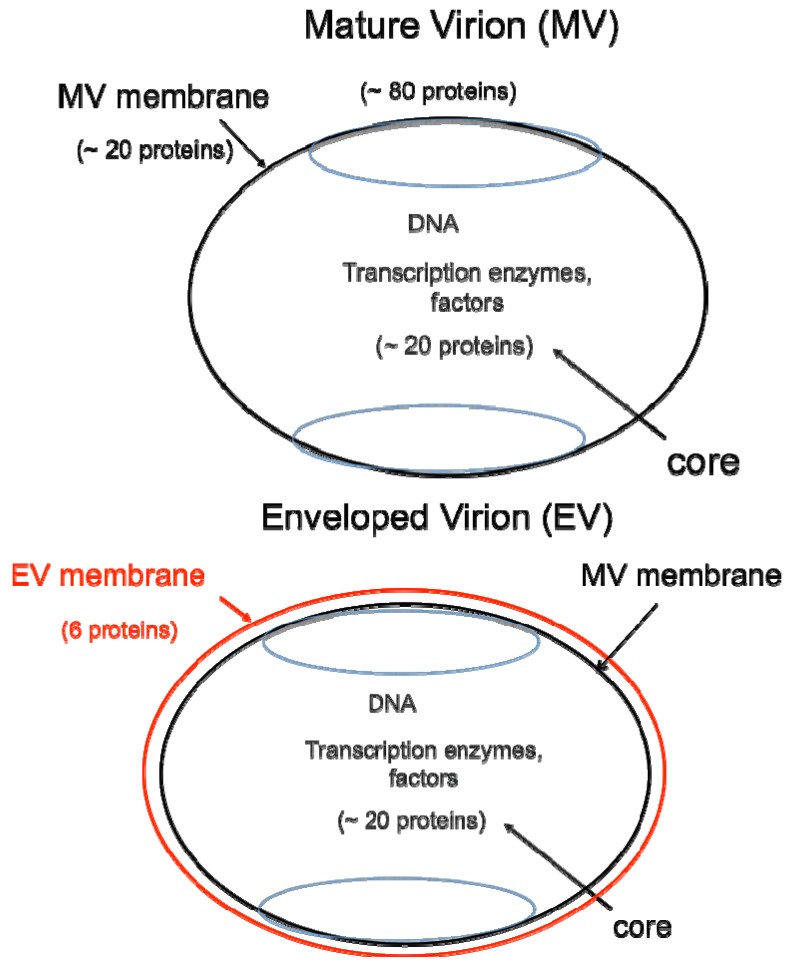
For many viruses, one or two proteins enable cell binding, membrane fusion and entry. The large number of proteins employed by poxviruses is unprecedented and may be related to their ability to infect a wide range of cells. There are two main infectious forms of vaccinia virus, the prototype poxvirus: the mature virion (MV), which has a single membrane, and the extracellular enveloped virion (EV), which has an additional outer membrane that is disrupted prior to fusion. Four viral proteins associated with the MV membrane facilitate attachment by binding to glycosaminoglycans or laminin on the cell surface, whereas EV attachment proteins have not yet been identified. Entry can occur at the plasma membrane or in acidified endosomes following macropinocytosis and involves actin dynamics and cell signaling. Regardless of the pathway or whether the MV or EV mediates infection, fusion is dependent on 11 to 12 non-glycosylated, transmembrane proteins ranging in size from 4- to 43-kDa that are associated in a complex. These proteins are conserved in poxviruses making it likely that a common entry mechanism exists. Biochemical studies support a two-step process in which lipid mixing of viral and cellular membranes is followed by pore expansion and core penetration.

Keywords: endocytosis; macropinocytosis; transmembrane proteins; vaccinia virus entry; viral membrane fusion.


Figures

Figure 1
Figure 1
Two major forms of infectious virions. The mature virion (MV) contains more than 80 proteins and consists of a nucleoprotein core surrounded by a lipid membrane (black) with about 20 proteins. Approximately 20 proteins within the core are devoted to synthesis and modification of mRNA. The enveloped virion (EV) consists essentially of a MV with an additional membrane (red) containing about 6 proteins distinct from those in the MV membrane.
Figure 2
Figure 2
Transmission electron micrographs showing a VACV MV fusing with the plasma membrane (A) and endosomal membrane (B). Prior to cryosectioning, the infected cells were stained with a monoclonal antibody to the MV membrane protein D8 followed by protein A conjugated to gold spheres. Modified from reference [25].
Figure 3
Figure 3
Effects of low pH and inhibitors of endosomal acidification on VACV MV entry. (A) Following adsorption of a recombinant VACV MV that expresses firefly luciferase at 4 °C, cells were incubated with buffers at the indicated pH for 3 min at 37 °C. The cells were then incubated with regular medium for 1 h at 37 °C, lysed and luciferase activity measured; (B) Cells were pretreated with indicated concentrations of bafilomycin A1 for 50 min at 37 °C and then infected with MVs, briefly exposed to pH 5 or 7.4 buffer, and luciferase activity measured as in panel A. Note that incubation at pH 5 allows fusion at the plasma membrane and bypasses the effect of the inhibitor. Modified from reference [25].
Figure 4
Figure 4
Membrane fusion and core entry. R18-loaded MVs that express firefly luciferase were incubated with HeLa cells at 4 °C to permit binding. Washed cells were then placed in a cuvette containing pre-warmed media at 37 °C and fluorescence was monitored over time (red line; left y-axis). In parallel, unlabeled MVs were bound to cells in the cold and then shifted to 37 °C. Cell lysates were prepared at indicated times and assayed for luciferase (LUC) activity (purple line; right y-axis). Modified from ref [107].

References

    1. Moss B. Poxviridae: The Viruses and Their Replication. In: Knipe D.M., Howley P.M., editors. Fields Virology. Vol. 2. Lippincott Williams & Wilkins; Philadelphia, PA, USA: 2007. pp. 2905–2946.
    1. Upton C., Slack S., Hunter A.L., Ehlers A., Roper R.L. Poxvirus orthologous clusters: Toward defining the minimum essential poxvirus genome. J. Virol. 2003;77:7590–7600. - PMC - PubMed
    1. Moss B. Poxvirus entry and membrane fusion. Virology. 2006;344:48–54. - PubMed
    1. Moss B., de Silva F. Poxvirus DNA Replication and Human Disease. In: DePamphilis M.L., editor. DNA Replication & Human Disease. Cold Spring Harbor Laboratory Press; Cold Spring Harbor, NY, USA: 2006. pp. 707–727.
    1. Yang Z., Reynolds S.E., Martens C.A., Bruno D.P., Porcella S.F., Moss B. Expression profiling of the intermediate and late stages of poxvirus replication. J. Virol. 2011;85:9899–9908. - PMC - PubMed

Publication types

LinkOut - more resources