Review
. 2012 May;4(5):800-32.
doi: 10.3390/v4050800. Epub 2012 May 10.

Herpes virus fusion and entry: a story with many characters

Affiliations
Review

Herpes virus fusion and entry: a story with many characters

Roselyn J Eisenberg et al. Viruses. 2012 May.

Abstract

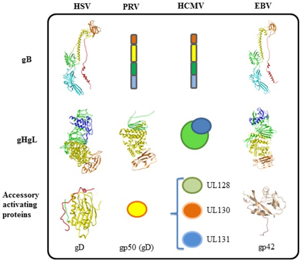
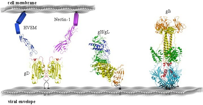
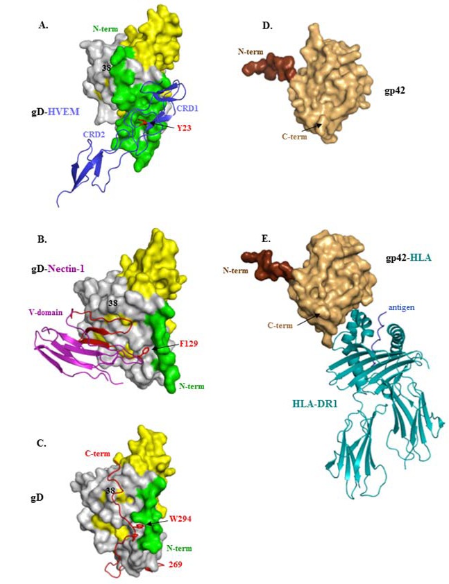
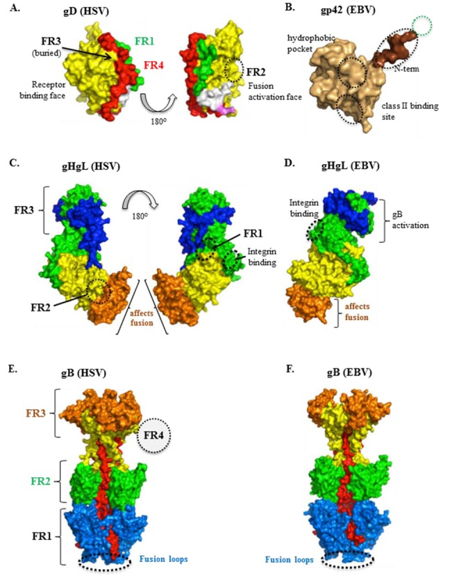
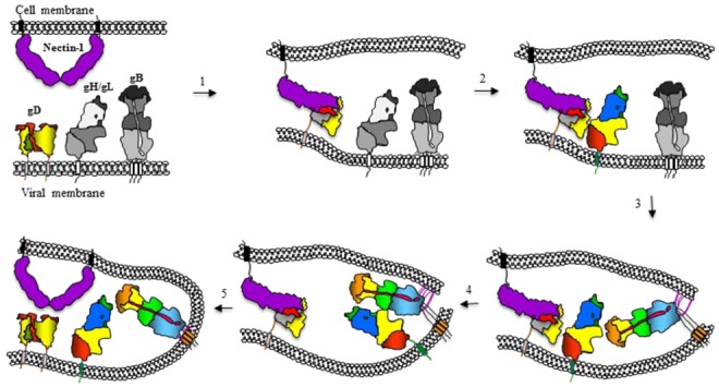
Herpesviridae comprise a large family of enveloped DNA viruses all of whom employ orthologs of the same three glycoproteins, gB, gH and gL. Additionally, herpesviruses often employ accessory proteins to bind receptors and/or bind the heterodimer gH/gL or even to determine cell tropism. Sorting out how these proteins function has been resolved to a large extent by structural biology coupled with supporting biochemical and biologic evidence. Together with the G protein of vesicular stomatitis virus, gB is a charter member of the Class III fusion proteins. Unlike VSV G, gB only functions when partnered with gH/gL. However, gH/gL does not resemble any known viral fusion protein and there is evidence that its function is to upregulate the fusogenic activity of gB. In the case of herpes simplex virus, gH/gL itself is upregulated into an active state by the conformational change that occurs when gD, the receptor binding protein, binds one of its receptors. In this review we focus primarily on prototypes of the three subfamilies of herpesviruses. We will present our model for how herpes simplex virus (HSV) regulates fusion in series of highly regulated steps. Our model highlights what is known and also provides a framework to address mechanistic questions about fusion by HSV and herpesviruses in general.

Keywords: CMV; EBV; HSV; VZV; crystal structure; functional region; glycoproteins; monoclonal antibody.


Figures

Figure 1
Figure 1
Fusion machinery of herpesviruses. All herpesviruses utilize gB and gHgL for membrane fusion. Different herpesviruses have different accessory proteins involved in regulation of membrane fusion. gD is used by alpha herpes viruses (HSV), and porcine herpesvirus (PRV) uses gp50. Beta herpesviruses (HCMV) use UL128, UL130, and UL131. Gamma herpesviruses (EBV) use accessory protein gp42. The only structures that are currently known can be accessed from the PDB database: HSV-1 gB, 2GUM; HSV-2 gHgL, 3M1C; HSV-1 gD, 2C36; EBV gB, 3FVC; EBV gHgL, 3PHF; EBV gp42, 3FD4; PRV gH, 2XQY.
Figure 2
Figure 2
Fusion machinery of HSV. The minimal set of proteins required for HSV membrane fusion is depicted to scale. gB is structurally conserved across all herpesviruses, while gH/gL is more variable. gD is the accessory fusion protein required for membrane fusion in HSV. gD determines cell tropism by binding cellular receptors HVEM or Nectin-1. All proteins are colored according to their structural regions, as defined previously for HSV gD, HSV gH/gL, HSV gB, HVEM and Nectin-1.
Figure 3
Figure 3
Structures of HSV gD and EBV gp42 alone or bound to their ligands. (A) gD(285t) bound to HVEM. The gD Ig fold is shown in yellow and its core extensions are in gray. The N-terminus (aa 1-32 in green) forms a hairpin bent at residue 21. The first two cysteine-rich domains (CRD) of HVEM are shown as blue ribbons. The essential tyrosine 23, which inserts between the strands of the gD N-terminal hairpin, is shown in red; (B) gD(285t) bound to nectin-1. gD is colored as in A. The N-terminal and C-terminal flexible residues (1-22, 260-285) are not solved. The V-domain of nectin-1 is shown as blue ribbons with beta strands CC’C” that contact gD shown in red. The essential phenylalanine 129, which inserts into a pocket near the N-terminus of gD is colored red; (C) Unbound HSV gD306t(307Cys) protomer. gD is colored as in A with the C-terminus (269-307) shown as a red ribbon. The C-terminus anchoring residue tryptophane 294 is shown in red and the position of Tyrosine 38 is indicated. The extended N-terminal 22 residues and the flexible hinge (aa. 260-268) are not visible; (D) Unbound EBV gp42 protein is shown in tan and residues at the N-terminus of the solved structure are colored brown (aa 83-93); (E) gp42 bound to human HLA protein. gp42 is colored as in D. The human HLA-DR1 is shown as teal ribbons and the antigenic peptide is colored blue. In all panels N and C-terminal residues of gD and gp42 are shown when visible. Representations of gD and gp42 are not drawn to the same scale.
Figure 4
Figure 4
Functional regions in herpes glycoproteins. All proteins are colored according to their structural regions, as defined previously for HSV gD, HSV gH/gL, and HSV gB. (A) HSV gD. Two views of the monomeric, unbound form of gD are shown. The green (part of which is FR1; 27-43) represents the N-terminus which forms the HVEM-binding hairpin in the gD-HVEM complex. The Ig-like core (contains FR2, residues 125-161, and FR3, residues 225-246) is shown in yellow and the C-terminus (contains FR4; 277-310) in red. For reference, the MC2 (234-250; white) and MC10 (262-272; pink) epitopes are shown; (B) EBV gp42. Defined regions are as described in Kirschner et al. Binding sites for proteins gH/gL (residues 36-81, within the green dotted circle) and HLA class II are indicated, as well as a distinct hydrophobic pocket essential for membrane fusion (black circles). The last two regions were defined by linker insertions; (C) HSV gH/gL. Two views of gH/gL are shown. The gH structural domains are H1 (green), H2 (yellow), and H3 (orange). gL is shown in blue. HSV gH/gL FRs are defined by the epitopes of neutralizing MAbs: LP11 (FR1), 52S (FR2), and CHL17/32 (FR3). The integrin binding site (residues 176-178) is also indicated. Numerous mutations that affect cell-cell fusion map to domain H3; it is currently unknown if this is a functional domain separate from FR2 (which spans H2-H3); no known neutralizing MAbs map here; (D) EBV gH/gL. gL (blue) contains a hypothesized site of gB interaction. The integrin binding site (residues 188-190) is also indicated. Numerous mutations that affect fusion map to domain H3, as well as the epitope of the neutralizing MAb CL 59; (E) HSV gB. HSV gB FRs are defined by the epitopes of neutralizing MAbs: SS55/SS106/SS144 (FR1, blue and red, fusion domain), C226/H1838/H1781 (FR2, green, gH/gL interaction domain), SS10/SS67-69 (FR3, orange, receptor-binding domain). The epitope of MAb H1817 (FR4, dotted circle) maps to the unresolved N-terminus. The fusion loops are indicated at the bottom of the structure; (F) EBV gB. Chimeric EBV mutants have suggested that structural domains III (yellow), IV (orange) and V (red) are important for gB-gH/gL binding. The fusion loops are indicated at the bottom of the structure.
Figure 5
Figure 5
Cartoon representation of the sequential events leading to HSV entry. The entry process begins with the binding of gD to specific receptors (nectin-1 is shown), to gain entry into target cells (step 1). Receptor binding triggers displacement of the C-terminus of gD to expose a previously hidden region of gD, which we propose interacts with gH/gL (step 2). This interaction results in a conformational change in gH/gL that enables it to up-regulate gB into a fusogenic state. This three-step process may involve the interaction of gB with a cell surface protein, the insertion of gB fusion loops into the opposing lipid membrane (step 3) and an interaction between the ectodomains of gB and gH/gL (step 4). This converts gB from a pre- to a post-fusion state, resulting in fusion of the viral envelope with cell membranes and delivery of the nucleocapsid into the target cell (step 5). All essential proteins shown were drawn based on published structures with the corresponding domains. Where pre-activation structures were not available, the proteins are shown in gray.

Similar articles

Cited by

References

    1. Campadelli-Fiume G., Menotti L. Entry of Alphaherpesviruses into the Cell. In: Arvin A., Campadelli-Fiume G., Mocarski E., Moore P.S., Roizman B., Whitley R., Yamanishi K., editors. Source Human Herpesviruses: Biology, Therapy, and Immunoprophylaxis. Cambridge University Press; Cambridge, UK: 2007. Chapter 7. - PubMed
    1. Campadelli-Fiume G., Menotti L., Avitabile E., Gianni T. Viral and cellular contributions to herpes simplex virus entry into the cell. Curr. Opin. Virol. 2012;2:28–36. - PubMed
    1. Navaratnarajah C.K., Miest T.S., Carfi A., Cattaneo R. Targeted entry of enveloped viruses: Measles and herpes simplex virus I. Curr. Opin. Virol. 2012;2:43–49. - PMC - PubMed
    1. Connolly S.A., Jackson J.O., Jardetzky T.S., Longnecker R. Fusing structure and function: A structural view of the herpesvirus entry machinery. Nat. Rev. Microbiol. 2011;9:369–381. - PMC - PubMed
    1. Hutt-Fletcher L.M., Chesnokova L.S. Integrins as triggers of Epstein-Barr virus fusion and epithelial cell infection. Virulence. 2010;1:395–398. - PMC - PubMed

Publication types

LinkOut - more resources