Review
. 2012 Mar;2(3):120023.
doi: 10.1098/rsob.120023.

The overlooked greatwall: a new perspective on mitotic control

Affiliations
Review

The overlooked greatwall: a new perspective on mitotic control

David M Glover. Open Biol. 2012 Mar.

Abstract

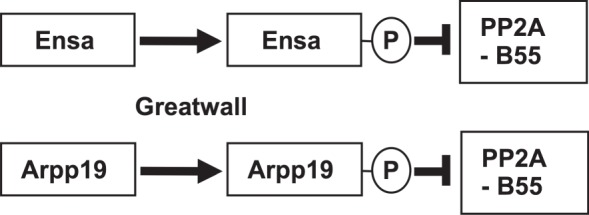
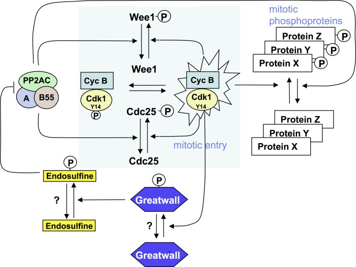
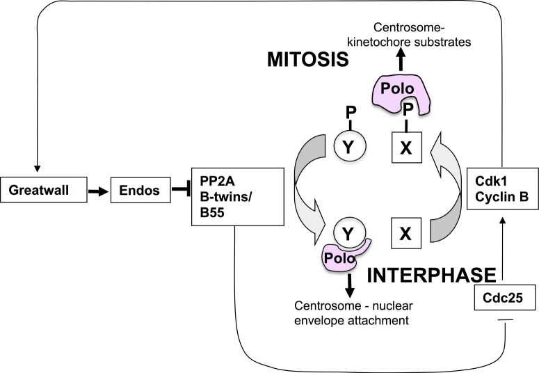
The role of the dual specificity protein phosphatase, Cdc25, in activating the cyclin-dependent kinase-cyclin B complex (Cdk1-CycB) by overcoming the inhibitory Wee1 kinase is a long-established principle for mitotic entry. Recently, however, evidence has emerged of a regulatory network that facilitates Cdk1-CycB activity by inhibiting the form of protein phosphatase 2A having a B55 regulatory subunit (PP2A-B55). Here, I review the genetic and biochemical evidence for Greatwall kinase and its substrate Endosulphine as the key components of this previously obscure regulatory network. Not only is the inhibition of PP2A-B55 by phospho-endosulphine required to prevent dephosphorylation of Cdk1-CycB substrates until mitotic exit, but it is also required to promote Cdc25 activity and inhibit Wee1 at mitotic entry. I discuss how these alternating states of preferential PP2A-B55 or Cdk1-CycB activity can have an impact upon the regulation of Polo kinase and its ability to bind different partner proteins as mitosis progresses.

Keywords: Endos; Greatwall kinase; PP2A; mitosis.


Figures

Figure 1.
Figure 1.
Origins of some of the alleles of polo and greatwall. (a) The orginal polo allele was identified as a mutation that, when homozygous in the mother, led to mitotic abnormalities and death of syncytial Drosophila embryos. Scant was isolated as a mutation that, when trans-heterozygous with polo (one mutant copy of Scant and one mutant copy of polo), caused females to produce embryos that died owing to a specific mitotic defect—loss of centrosomes from one pole (see text and figure 2). Scheme for the identification of mutations that would suppress the + Scant/polo + phenotype. These were polo+ duplications; revertants of Scant to its recessive alleles, Sr; and a second-site suppressor, su. Sr was identified as greatwall, su as endos. We may also refer to Scant as gwlScant.
Figure 2.
Figure 2.
The ‘Scant’ phenotype; its suppressor and enhancers. Typical mitotic figure from embryos derived from a mother with one mutant copy of polo and one mutant copy of gwlScant. We first named the gene Scant after Scott of the Antarctic, the British explorer who set out to find the mysterious southern geomagnetic pole of the Earth in a ship, the Discovery, which is now anchored in full view of the University of Dundee campus. Reducing the wild-type gene dosage of endos in the mother suppresses, whereas increasing the endos gene dosage enhances the phenotype.
Figure 3.
Figure 3.
Suppression of the endos knockdown phenotype in cultured cells by simultaneous knockdown of PP2A-B55twins. (a) RNAi-mediated depletion of either Greatwall or Endos results in prolonged mitoses in which chromosomes remain scattered on elongated spindles before attempting anaphase. This phenotype is suppressed by the simultaneous depletion of either the catalytic subunit of PP2A (encoded by mts—microtubule star), the structural A subunit (encoded by PP2A 29B) or the B subunit (encoded by twins also known as aar-abnormal anaphase resolved). It is not suppressed by knocking down the three other regulatory B subunits in Drosophila (wdb,widerborst; B′; or B″). (b) Cells depleted of Endos display lagging chromosomes at anaphase. This phenotype is rescued by simultaneous depletion of the PP2A B-subunit, Twins.
Figure 4.
Figure 4.
The paralogues Ensa (Endos) and Arpp19 are phosphorylated by Greatwall kinase to become inhibitors of PP2A-B55.
Figure 5.
Figure 5.
Greatwall–Endos regulates mitotic entry and stabilizes the mitotic state by inhibiting PP2A-B55. Mitotic entry is regulated by a positive-amplification loop in which the dual-specificity phosphatase Cdc25 dephosphorylates and thereby activates Cdk1-cycB kinase. Cdk1-cycB phosphorylates and activates Cdc25. Cdc25 is opposed by the Wee1 kinase that is inhibited by Cdk1-cycB phosphorylation. Thus, PP2A dephosphorylates Cdc25-P and Wee1-P to oppose Cdk1-cycB. This accounts for the long-known fact that inhibition of PP2A (by okadaic acid) promotes mitotic entry. Endos, phosphorylated by Greatwall, acts in an analogous way. By inhibiting PP2A, phospho-Endos also maintains the mitotic state by enabling the multiple mitotic substrates of Cdk1-cycB to retain their phosphate groups.
Figure 6.
Figure 6.
Hypothesis for how Greatwall might act antagonistically to Polo late in mitosis in the syncytial nuclear division cycles of the Drosophila embryo. As both Greatwall and Polo are ‘mitotic kinases’, it seems counterintuitive that Greatwall might inhibit some Polo functions as suggested by the interactions between the gwlScant and polo mutations. Several explanations for this are possible and are discussed in the text. This schematic presents one of these potential explanations. It postulates that because Polo can interact in mitosis with proteins (X-P) that have been phosphorylated by Cdk1-cycB, and at mitotic exit and interphase with proteins that do not have such mitotic phosphorylations (Y), downregulating PP2A in Greatwall-Scant-derived embryos can prolong Polo's interactions with its mitotic partners and deny its interactions with interphase partners. In the context of this scheme, the consequence of the latter would be to favour retention of high Y-P levels and thereby lead to loss of centrosomes from nuclei, postulated to be an interphase process requiring dephosphorylated protein Y.

Similar articles

Cited by

References

    1. Masui Y, Markert CL. 1971. Cytoplasmic control of nuclear behavior during meiotic maturation of frog oocytes. J. Exp. Zool. 177, 129–14510.1002/jez.1401770202 (doi:10.1002/jez.1401770202) - DOI - DOI - PubMed
    1. Reynhout JK, Smith LD. 1974. Studies on the appearance and nature of a maturation-inducing factor in the cytoplasm of amphibian oocytes exposed to progesterone. Dev. Biol. 38, 394–40010.1016/0012-1606(74)90016-5 (doi:10.1016/0012-1606(74)90016-5) - DOI - DOI - PubMed
    1. Kishimoto T, Kanatani H. 1976. Cytoplasmic factor responsible for germinal vesicle breakdown and meiotic maturation in starfish oocyte. Nature 260, 321–32210.1038/260321a0 (doi:10.1038/260321a0) - DOI - DOI - PubMed
    1. Guerrier P, Doree M, Freyssimet G. 1975. Early stimulation of protein kinase activity during hormonal meiosis reinitiation in starfish ovocytes. C R. Acad. Sci. Hebd. Seances Acad. Sci. D. 281, 1475–1478 - PubMed
    1. Lohka M, Masui Y. 1983. Formation in vitro of sperm pronuclei and mitotic chromosomes induced by amphibian ooplasmic components. Science 220, 719–72110.1126/science.6601299 (doi:10.1126/science.6601299) - DOI - DOI - PubMed

Publication types

LinkOut - more resources