. 2012 Mar;2(3):120030.
doi: 10.1098/rsob.120030.

Initiating head development in mouse embryos: integrating signalling and transcriptional activity

Affiliations

Initiating head development in mouse embryos: integrating signalling and transcriptional activity

Ruth M Arkell et al. Open Biol. 2012 Mar.

Abstract

The generation of an embryonic body plan is the outcome of inductive interactions between the progenitor tissues that underpin their specification, regionalization and morphogenesis. The intercellular signalling activity driving these processes is deployed in a time- and site-specific manner, and the signal strength must be precisely controlled. Receptor and ligand functions are modulated by secreted antagonists to impose a dynamic pattern of globally controlled and locally graded signals onto the tissues of early post-implantation mouse embryo. In response to the WNT, Nodal and Bone Morphogenetic Protein (BMP) signalling cascades, the embryo acquires its body plan, which manifests as differences in the developmental fate of cells located at different positions in the anterior-posterior body axis. The initial formation of the anterior (head) structures in the mouse embryo is critically dependent on the morphogenetic activity emanating from two signalling centres that are juxtaposed with the progenitor tissues of the head. A common property of these centres is that they are the source of antagonistic factors and the hub of transcriptional activities that negatively modulate the function of WNT, Nodal and BMP signalling cascades. These events generate the scaffold of the embryonic head by the early-somite stage of development. Beyond this, additional tissue interactions continue to support the growth, regionalization, differentiation and morphogenesis required for the elaboration of the structure recognizable as the embryonic head.

Keywords: gene transcription; head formation; morphogenesis; mouse embryo; signalling.


Figures

Figure 1.
Figure 1.
Development of the mouse embryo from 3.5 dpc (days post coitum) to 6.5 dpc. (a) Blastocyst containing an inner cell mass comprising the epiblast and the primitive endoderm. (b,c) Egg cylinder embryo at 5.0 dpc with distal visceral endoderm, and 5.5 dpc with anterior visceral endoderm. (d) Early-streak embryo at 6.5 dpc, with formation of the primitive streak and the nascent mesoderm.
Figure 2.
Figure 2.
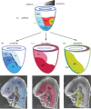
Allocation of the germ layer derivatives to the embryonic head structures. (a) Regionalization of germ layer progenitors in the epiblast elicited by the graded signalling activity across the prospective anterior–posterior plane of the embryo. (b–d) Allocation of epiblast-derived cells during gastrulation to (b) the ectoderm tissues that contribute to the brain, neural crest and the surface ectoderm, (c) the mesoderm tissues in the cranial mesenchyme and the heart, and (d) endoderm tissues of the embryonic foregut. The fate maps of the progenitor tissues of the embryonic head reveals that the domains and boundaries of the progenitors in the three germ layers are generally aligned with each other, although a clear demarcation of head versus non-head progenitors is not yet evident at the late gastrulation stage. ade, anterior definitive (gut) endoderm; ame, anterior mesendoderm; amn, amnion ectoderm; ave, anterior visceral endoderm; crm, cranial mesoderm; en, endoderm; fb, forebrain; fg, foregut; hb, hindbrain; ht, heart; md, midbrain; mes, mesoderm; ncc, neural crest cells; n-ect, neuroectoderm; se, surface ectoderm.
Figure 3.
Figure 3.
Inductive interaction between the germ layer derivatives during head formation. (a) A schematic diagram of the right-hand half of the late-streak embryo showing the domains of brain progenitors in the ectoderm and the opposing paraxial mesoderm, endoderm and the axial mesendoderm (prechordal plate and anterior notochord, derived from the node). The boxed area of (a) is shown in (b), which depicts the planar (inductive and suppressive) interaction between the prechordal plate and the anterior notochord, the induction by the prechordal plate to maintain the anterior definitive endoderm, and the vertical (i.e. between germ layers) induction of the neural primordium by the endoderm and the axial mesendoderm.
Figure 4.
Figure 4.
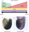
Specification of the brain progenitors is facilitated by WNT signalling activity. (a) The regionalized activity of signalling components sets up a signalling landscape with (b) reduced WNT signal activity (low reporter expression) in (c) the domain of brain progenitor (marked by Otx2 expression). Source of figures: (a) fig. 9, Fossat et al. [44]; (b) fig. 1c, Lewis et al. [43] (permission for use by authors under copyright agreement with Development, Company of Biologists Ltd).
Figure 5.
Figure 5.
The intersection of signalling and transcriptional activity culminates in the suppression of BMP, WNT and Nodal signalling for head formation. Sources of signals in the gastrulation stage embryo: Bone Morphogenetic Protein (BMP) from the extraembryonic ectoderm and the posterior epiblast, and WNT (Wnt2b, Wnt3, Wnt3a and Wnt8a) and Nodal from the posterior epiblast and primitive streak. BMP antagonists Noggin (Nog) and Chordin (Chrd); Nodal antagonist Lefty1; WNT antagonists Dickkorf-1 (Dkk1) and Secreted frizzled-related proteins (Sfrps). Transcription factors: Goosecoid (Gsc), LIM homeobox protein 1 (Lhx1), LIM domain binding 1 (Ldb1), Single-stranded DNA binding protein 1 (Ssdp1); Orthodenticle homologue 2 (Otx2), SRY-box containing gene 17 (Sox17).

Similar articles

Cited by

References

    1. Rossant J, Tam PP. 2004. Emerging asymmetry and embryonic patterning in early mouse development. Dev. Cell 7, 155–16410.1016/j.devcel.2004.07.012 (doi:10.1016/j.devcel.2004.07.012) - DOI - DOI - PubMed
    1. Tam PP, Gad JM. 2004. Gastrulation of the mouse embryo. In Gastrulation (ed. Stern C. D.), pp. 223–262 Cold Spring Harbor, NY: Cold Spring Harbour Laboratory Press
    1. Tam PP, Loebel DA. 2007. Gene function in mouse embryogenesis: get set for gastrulation. Nat. Rev. Genet. 8, 368–38110.1038/nrg2084 (doi:10.1038/nrg2084) - DOI - DOI - PubMed
    1. Tam PP. 1989. Regionalisation of the mouse embryonic ectoderm: allocation of prospective ectodermal tissues during gastrulation. Development 107, 55–67 - PubMed
    1. Cajal M, Lawson KA, Hill B, Moreau A, Rao J, Ross A, Collignon J, Camus A. 2012. Clonal and molecular analysis of the prospective anterior neural boundary in the mouse embryo. Development 139, 423–43610.1242/dev.075499 (doi:10.1242/dev.075499) - DOI - DOI - PMC - PubMed

Publication types

LinkOut - more resources