. 2012 Jun 1;4(3):272-83.
doi: 10.3978/j.issn.2072-1439.2012.05.07.

Clinicopathologic analysis of cardiac myxomas: Seven years' experience with 61 patients

Clinicopathologic analysis of cardiac myxomas: Seven years' experience with 61 patients

Ji-Gang Wang et al. J Thorac Dis. .

Abstract

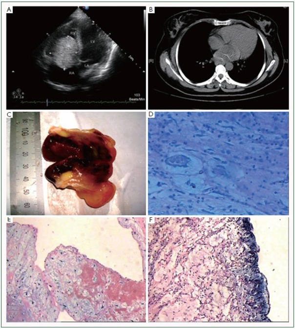
Objective: Cardiac myxomas are the most common primary neoplasms of heart. The present study was performed on the 61 cases of patients with cardiac myxoma, in order to investigate the tumors' clinical and pathological features, and to identify the relationship between the pathological characteristics and clinical behaviors.

Methods: A total of 61 cardiac myxoma cases were analyzed and reviewed retrospectively, including the clinical presentations, physical examinations, and echocardiography, electrocardiography, and pathology documents.

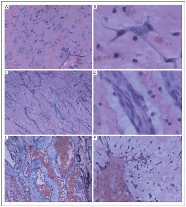
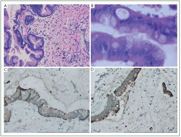
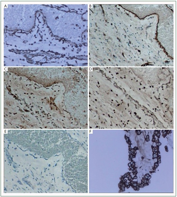
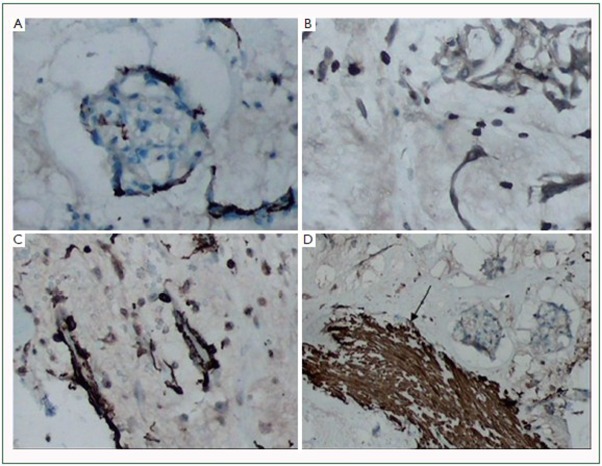
Results: The total patient cohort was made up of 37 women and 24 men. The average age at diagnosis was 48.8 years in males and 51.9 years in females. The most common complaint was dyspnea (37 cases, 60.7%) and the most common sign was systolic murmur (30 cases, 49.2%). Two surface structures and three tumor cell arrangement patterns were observed, and statistical analysis revealed the surface structure was related to the cell arrangement pattern. However, neither the cell arrangement pattern nor the tumor surface structure showed a significant correlation with the clinical presentation.

Conclusions: The present study showed the pathological profiles of cardiac myxomas were not related to the clinical presentations. The results of our study indicate morphologic classifications of cardiac myxomas may not be significant for clinical practice.

Keywords: Cardiac neoplasms; immunohistochemistry; myxoma; neoplasm recurrence, local; pathology, surgical.


Figures

Figure 1
Figure 1
Echocardiography, computed tomography, gross appearance, AB-PAS staining, and surface appearance of myxomas. A: echocardiography could well identify a right ventricle myxoma; B: computed tomography showed a low density mass located in the left atrium; C: on gross examination, the tumors often have a pedicle. The tumor mass is soft, gelatinous, and very friable; D: AB-PAS staining showed the stromal matrix appeared to purplish red (positive for AB and PAS), while the mucous halo showed blue color (positive for AB); E: the tumor surface showed a papillary appearance (H&E staining); F: the tumor surface showed a smooth and solid appearance (H&E staining). (original magnifications: D, ×100; E, F, ×40).
Figure 2
Figure 2
Three cell arrangement subtypes of myxomas. A, B: single cell predominant subtype; the single myxoid cells were scattered; C, D: cell cord predominant subtype; myxomas of this type showed a rudimentary “chicken-wire” vessels like appearance, with few and scattered single myxoid cells in the stroma; E, F: vasoformative ring predominant subtype; myxomas of this type showed a hemangioma-like appearance, containing distended blood sinus and distinct mucous halo. (H&E staining, original magnifications: A, C, E, ×40; B, D, ×400; F, ×200).
Figure 3
Figure 3
Typical glandular structures in myxomas. A, B: Well-defined mucous glands, containing goblet cells interspersed between cells, formed by columnar epithelium were observed (H&E staining); C, D: the glandular structures bear positive expression of CK and EMA. (original magnifications: A, C, D, ×100; B, ×400).
Figure 4
Figure 4
Immunohistochemical staining for Vimentin, CD34, CD68 and SMA in myxomas. A: Vimentin was diffusely and strongly reactive with all tumor cells and blood vessels; B: CD34 staining; C: SMA staining; D: CD68 staining; E: negative control. We observed that SMA, CD34 and CD68 were expressed variably in myxomas; F: the surface lining cells were immunoreactive for CD34. (original magnifications: A, B, C, D, E, ×40; F, ×100).
Figure 5
Figure 5
Immunohistochemical staining for CD34, CD68 and SMA in myxomas. A: CD34; B: CD68; C, D: SMA. Our immunostaining results showed only part of tumor cells showed positive immunoreactivity for these antibodies. Note the arrow in figure 5D, smooth muscle of blood vessel bear strong immunoreactivity for SMA. (original magnifications: A, ×200; B, C, D, ×100).

Similar articles

Cited by

References

    1. Wold LE, Lie JT. Cardiac myxomas: a clinicopathologic profile. Am J Pathol. 1980;101:219-240 - PMC - PubMed
    1. Burke AP, Tazelaar H, Gomez-Roman JJ, et al. Cardiac myxoma. In: Travis WD, Brambilla E, Muller-Hermelink HK, Harris CC, editors. Pathology and Genetics of Tumours of the Lung, Pleura, Thymus and Heart. Lyon: IARC Press, 2004: 260-3.
    1. Lie JT. The identity and histogenesis of cardiac myxomas: a controversy put to rest. Arch Pathol Lab Med. 1989;113:724-726 - PubMed
    1. Burke AP, Virmani R. Cardiac myxoma. A clinicopathologic study. Am J Clin Pathol. 1993;100:671-680 - PubMed
    1. Kodama H, Hirotani T, Suzuki Y, et al. Cardiomyogenic differentiation in cardiac myxoma expressing lineage-specific transcription factors. Am J Pathol. 2002;161:381-389 - PMC - PubMed