Asian sand dust enhances allergen-induced th2 allergic inflammatory changes and mucin production in BALB/c mouse lungs
- PMID: 22754714
- PMCID: PMC3378927
- DOI: 10.4168/aair.2012.4.4.206
Asian sand dust enhances allergen-induced th2 allergic inflammatory changes and mucin production in BALB/c mouse lungs
Abstract
Purpose: Recent studies have reported that Asian sand dust (ASD) has a potential risk of aggravating airway inflammation. The purpose of this study was to investigate the effect of ASD on inflammation and mucin production in the airways of allergic mice.
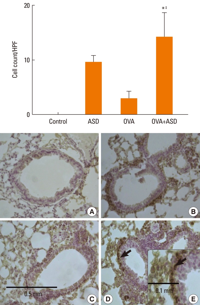
Methods: FORTY BALB/C FEMALE MICE WERE DIVIDED INTO FOUR GROUPS: saline (group 1); ASD (group 2); ovalbumin (OVA) alone (group 3); and OVA+ASD (group 4). OVA-specific immunoglobulin E (IgE) in serum and interleukin (IL)-4, IL-5, IL-13, and interferon-gamma (IFN-γ) in bronchoalveolar lavage fluid (BALF) were measured by enzyme-linked immunosorbent assay (ELISA). Hematoxylin & eosin (H&E) and Periodic acid-Schiff (PAS) staining was performed on lung tissues. In addition, immunohistochemical staining for IL-4, IL-5, MUC5AC, and transforming growth factor alpha (TGF-α) was conducted.
Results: Serum IgE levels were significantly higher in group 4 than in group 3 (P<0.05). IL-4 and IL-5 in BALF were significantly higher in group 4 than in group 3 (P<0.05, respectively). Based on H&E staining, inflammatory cell numbers were significantly greater in group 4 than in the other groups (P<0.05). The number of PAS-positive cells was also significantly greater in groups 3 and 4 than in groups 1 and 2 (P<0.05). The numbers of IL-4 and IL-5-positive cells were higher in group 4 than in group 3 (P<0.05). The number of MUC5AC and TGF-α-positive cells were also higher in group 4 than in group 3 (P<0.05).
Conclusions: Our data suggest that ASD increases cytokine expression and mucin production in an allergic murine model. The increased inflammatory reactions were related to cytokine production.
Keywords: Asian sand dust; airway inflammation; mucin.
Conflict of interest statement
There are no financial or other issues that might lead to conflict of interest.
Figures


Similar articles
-
Effect of Asian sand dust on mucin production in NCI-H292 cells and allergic murine model.Otolaryngol Head Neck Surg. 2012 Jun;146(6):887-94. doi: 10.1177/0194599812439011. Epub 2012 Mar 8. Otolaryngol Head Neck Surg. 2012. PMID: 22402586
-
Enhancement of OVA-induced murine lung eosinophilia by co-exposure to contamination levels of LPS in Asian sand dust and heated dust.Allergy Asthma Clin Immunol. 2014 Jun 9;10(1):30. doi: 10.1186/1710-1492-10-30. eCollection 2014. Allergy Asthma Clin Immunol. 2014. PMID: 24982682 Free PMC article.
-
Co-exposure of peptidoglycan and heat-inactivated Asian sand dust exacerbates ovalbumin-induced allergic airway inflammation in mice.Inhal Toxicol. 2022;34(9-10):231-243. doi: 10.1080/08958378.2022.2086650. Epub 2022 Jun 13. Inhal Toxicol. 2022. PMID: 35698289
-
Comparison of asthma phenotypes in OVA-induced mice challenged via inhaled and intranasal routes.BMC Pulm Med. 2019 Dec 10;19(1):241. doi: 10.1186/s12890-019-1001-9. BMC Pulm Med. 2019. PMID: 31823765 Free PMC article.
-
Therapeutic potential of anti-IL-1β IgY in guinea pigs with allergic asthma induced by ovalbumin.Mol Immunol. 2014 Mar;58(1):139-49. doi: 10.1016/j.molimm.2013.11.006. Epub 2013 Dec 24. Mol Immunol. 2014. PMID: 24355520
Cited by
-
Titanium Dioxide Nanoparticle Exposure Provokes Greater Lung Inflammation in Females Than Males in the Context of Obesity.Int J Nanomedicine. 2025 Apr 24;20:5321-5336. doi: 10.2147/IJN.S508676. eCollection 2025. Int J Nanomedicine. 2025. PMID: 40297406 Free PMC article.
-
Mechanisms underlying the health effects of desert sand dust.Environ Int. 2021 Dec;157:106790. doi: 10.1016/j.envint.2021.106790. Epub 2021 Jul 29. Environ Int. 2021. PMID: 34333291 Free PMC article. Review.
-
Silicon Dioxide Nanoparticles Enhance Endotoxin-Induced Lung Injury in Mice.Molecules. 2018 Sep 3;23(9):2247. doi: 10.3390/molecules23092247. Molecules. 2018. PMID: 30177658 Free PMC article.
-
Asian Sand Dust Enhances the Inflammatory Response and Mucin Gene Expression in the Middle Ear.Clin Exp Otorhinolaryngol. 2016 Sep;9(3):198-205. doi: 10.21053/ceo.2015.01060. Epub 2016 Apr 20. Clin Exp Otorhinolaryngol. 2016. PMID: 27095518 Free PMC article.
-
The effects of BRL-50481 on ovalbumin-induced asthmatic lung inflammation exacerbated by co-exposure to Asian sand dust in the murine model.Arch Pharm Res. 2022 Jan;45(1):51-62. doi: 10.1007/s12272-021-01367-x. Epub 2022 Jan 4. Arch Pharm Res. 2022. PMID: 34984603 Free PMC article.
References
-
- Salvi S. Pollution and allergic airways disease. Curr Opin Allergy Clin Immunol. 2001;1:35–41. - PubMed
-
- Kim YK, Song SK, Lee HW, Kim CH, Oh IB, Moon YS, Shon ZH. Characteristics of Asian dust transport based on synoptic meteorological analysis over Korea. J Air Waste Manag Assoc. 2006;56:306–316. - PubMed
-
- Kang J, Choi MS, Lee CB. Atmospheric metal and phosphorus concentrations, inputs, and their biogeochemical significances in the Japan/East Sea. Sci Total Environ. 2009;407:2270–2284. - PubMed
-
- Duce RA, Unni CK, Ray BJ, Prospero JM, Merrill JT. Long-range atmospheric transport of soil dust from Asia to the tropical north pacific: temporal variability. Science. 1980;209:1522–1524. - PubMed
-
- Park SH, Song CB, Kim MC, Kwon SB, Lee KW. Study on size distribution of total aerosol and water-soluble ions during an Asian dust storm event at Jeju Island, Korea. Environ Monit Assess. 2004;93:157–183. - PubMed