

Routine Chest X-ray: Still Valuable for the Assessment of Left Ventricular Size and Function in the Era of Super Machines?
- PMID: 22754739
- PMCID: PMC3385501
- DOI: 10.4103/2156-7514.96540
Routine Chest X-ray: Still Valuable for the Assessment of Left Ventricular Size and Function in the Era of Super Machines?
Abstract
Objectives: The development of technologically advanced, expensive techniques has progressively reduced the value of chest X-ray in clinical practice for the assessment of left ventricular (LV) dilatation and dysfunction. Although controversial data are reported on the role of this widely available technique in cardiac assessment, it is known that the cardio-thoracic ratio is predictive of risk of progression in the NYHA Class, hospitalization, and outcome in patients with LV dysfunction. This study aimed to evaluate the reliability of the transverse diameter of heart shadow [TDH] by chest X-ray for detecting LV dilatation and dysfunction as compared to Magnetic Resonance Imaging (MRI) performed for different clinical reasons.
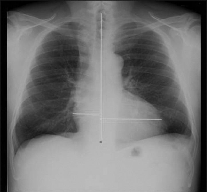
Materials and methods: In 101 patients, TDH was measured in digital chest X-ray and LV volumes and ejection fraction (EF) by MRI, both exams performed within 2 days.
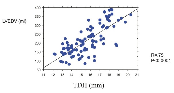
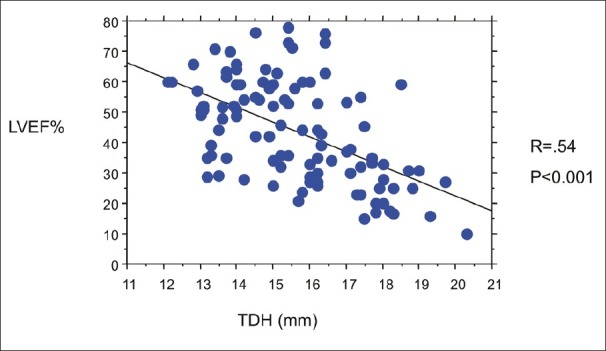
Results: A direct correlation between TDH and end-diastolic volumes (r = .75, P<0.0001) was reported. TDH cut-off values of 14.5 mm in females identified LV end-diastolic volumes >150 mL (sensitivity: 82%, specificity: 69%); in males a cut-off value of 15.5 mm identified LV end-diastolic volumes >210 mL (sensitivity: 84%; specificity: 72%). A negative relation was found between TDH and LVEF (r = -.54, P<0.0001). The above cut-off values of TDH discriminated patients with LV systolic dysfunction - LVEF <35% (sensitivity and specificity: 67% and 57% in females; 76% and 59% in males, respectively).
Conclusions: Chest X-ray may still be considered a reliable technique in predicting LV dilatation by the accurate measurement of TDH as compared to cardiac MRI. Technologically advanced, expensive, and less available imaging techniques should be performed on the basis of sound clinical requests.
Keywords: Cardiac MRI; chest X-ray; left ventricular dilatation; left ventricular dysfunction.
Conflict of interest statement
Figures
References
-
- Ferguson EC, Krishnamurthy R, Oldham SA. Classic imaging signs of cardiovascular abnormalities. Radiographics. 2007;27:1323–4. - PubMed
-
- Young JB. Imaging patients with heart failure: Expectations of the clinician. Heart Fail Clin. 2006;2:107–15. - PubMed
-
- Maffessanti M, Berlot G, Bortolotto P. Chest roentgenology in the intensive care unit: An overview. Eur Radiol. 1998;8:69–78. - PubMed
-
- Sanchez O, Revel MP, Couchon S, Meyer GL. Imaging of pulmonary hypertension. Rev Mal Resp. 2007;24:155–69. - PubMed
-
- Hunt SA, Abraham WT, Chin MH, Feldman AM, Francis GS, Ganiats TG, et al. ACC/AHA 2005 Guideline Update for the Diagnosis and Management of Chronic heart failure in the adult: A report of the American College of Cardiology/American Heart Association Task Force on Practice Guidelines (Writing Committee to Update the 2001 Guidelines for the Evaluation and Management of Heart Failure): Developed in Collaboration With the American College of Chest Physicians and the International Society for Heart and Lung Transplantation: Endorsed by the Heart Rhythm Society. Circulation. 2005;112:e154–235. - PubMed
LinkOut - more resources
Full Text Sources
