

CT Manifestations of Osler-Weber-Rendu Syndrome in Liver: Report of Three Cases
- PMID: 22754740
- PMCID: PMC3385500
- DOI: 10.4103/2156-7514.96541
CT Manifestations of Osler-Weber-Rendu Syndrome in Liver: Report of Three Cases
Abstract
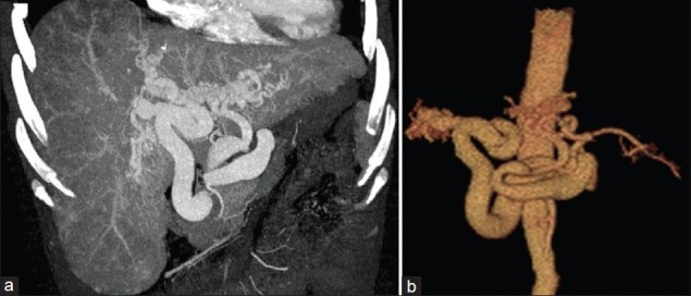
Osler-Weber-Rendu syndrome is characterized by widespread telangiectasias. Its clinical manifestations depend on position and scope of the abnormal vessels. The clinical and CT data of 3 patients with Osler-Weber-Rendu syndrome were retrospectively analyzed. CT features reviewed include the change of volume and configuration of liver, presence of tortuous and irregular vessels, opacified vessel mass, arteriovenous shunt, and splenomegaly. CT is helpful for diagnosis, treatment, and follow-up of Osler-Weber-Rendu syndrome.
Keywords: Computed tomography; Osler-Weber-Rendu syndrome; liver; maximum intensity projection; volume rendering technique.
Conflict of interest statement
Figures


References
-
- Fuchizaki U, Miyamori H, Kitagawa S, Kaneko S, Kobayashi K. Hereditary Haemorrhagic Telangiectasia (Rendu-Osler-Weber Disease) Lancet. 2003;362:1490–4. - PubMed
-
- Haitjema T, Disch F, Overtoom TT, Westermann CJ, Lammers JW. Screening family members of patients with hereditary haemorrhagic telangiectasia. Am J Med. 1995;99:519–24. - PubMed
-
- Bernard G, Mion F, Henry L, Plauchu H, Paliard P. Hepatic involvement in hereditary hemorrhagic telangiectasia: Clinical, radiological and hemodynamic studies of 11 cases. Gastroenterology. 1993;105:482–7. - PubMed
-
- Piskorz MM, Waldbaum C, Volpacchio M, Sordá J. Liver involvement in hereditary hemorrhagic telangiectasia. Acta Gastroenterol Latinoam. 2011;41:225–9. - PubMed
Publication types
LinkOut - more resources
Full Text Sources
