Immune reactivity in psoriatic munro-saboureau microabscesses, stratum corneum and blood vessels
- PMID: 22754876
- PMCID: PMC3385361
- DOI: 10.4103/1947-2714.97204
Immune reactivity in psoriatic munro-saboureau microabscesses, stratum corneum and blood vessels
Abstract
Background: A characteristic feature of early active psoriatic lesions is the intraepidermal penetration of neutrophils, with attendant formation of Munro-Saboureau microabscesses. Previous immunofluorescence studies have shown reactivity of in vivo binding of stratum corneum antibodies (SCAs) within the Munro-Saboreau microabscesses in cases of psoriasis.
Aims: In our study, we aimed to investigate any correlation between the SCAs and the Munro-Saboureau microabscesses.
Materials and methods: We investigated 50 archival biopsies of psoriasis with Munro-Saboureau microabscesses, and attempted to confirm antibody colocalization within these microabcesses via immunohistochemistry staining. As controls, we utilized 50 skin biopsies from healthy patients undergoing esthetic plastic surgery procedures.
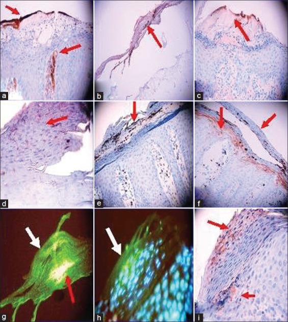
Results: Within the Munro-Saboureau microabscesses, the following markers were statistically significantly positive relative to controls: CD1a, CD8, CD23, cyclooxygenase-2, myeloid histoid antigen, albumin, fibrinogen, kappa, lambda, von Willebrand factor, IgG, IgM, IgD, complement/C3c, C3d, myeloperoxidase, and carcinoembryonic antigen (P < 0.05). Autoreactivity to blood vessels was also detected, with multiple immunoglobulins and complement factors.
Conclusions: We document important correlations between the Munro-Saboureau microabscesses, SCAs, and other immunoreactants.
Keywords: Autoimmunity; Blood vessels; Munro-Saboureau microabscesses carcinoembryonic antigen; Psoriasis; von Willebrand factor.
Conflict of interest statement
Figures
References
-
- Steffen C. William John Munro and Munro's abscess, and Franz Kogoj and Kogoj's spongiform pustule. Am J Dermatopathol. 2002;24:364–8. - PubMed
-
- Coimbra S, Figueiredo A, Castro E, Rocha-Pereira P, Santos-Silva A. The roles of cells and cytokines in the pathogenesis of psoriasis. Int J Dermatol. 2012;51:389–95. - PubMed
-
- Neumann E, Hård S. The significance of the epidermal sweat duct unit in the genesis of pustular psoriasis (Zumbusch) and the microabscess of Munro-Sabouraud. Acta Derm Venereol. 1974;54:141–6. - PubMed
-
- Dabski K, Glinski W, Beutner EH, Jablonska S. In vitro effects of enzymes of polymorphonuclear leukocytes on the antigenicity of stratum corneum. Int Arch Allergy Appl Immunol. 1985;77:287–91. - PubMed
-
- Kumar V, Jones P, Beutner EH, Jablonska S. Immunofluorescence studies in psoriasis: Detection of antibodies to stratum corneum in psoriatic scales. Ann NY Acad Sci. 1983;420:361–8. - PubMed
LinkOut - more resources
Full Text Sources
Research Materials