. 2013 Jan;37(1):96-109.
doi: 10.1111/j.1530-0277.2012.01850.x. Epub 2012 Jul 3.

Programmed cell death 4 (PDCD4): a novel player in ethanol-mediated suppression of protein translation in primary cortical neurons and developing cerebral cortex

Affiliations

Programmed cell death 4 (PDCD4): a novel player in ethanol-mediated suppression of protein translation in primary cortical neurons and developing cerebral cortex

Madhusudhanan Narasimhan et al. Alcohol Clin Exp Res. 2013 Jan.

Abstract

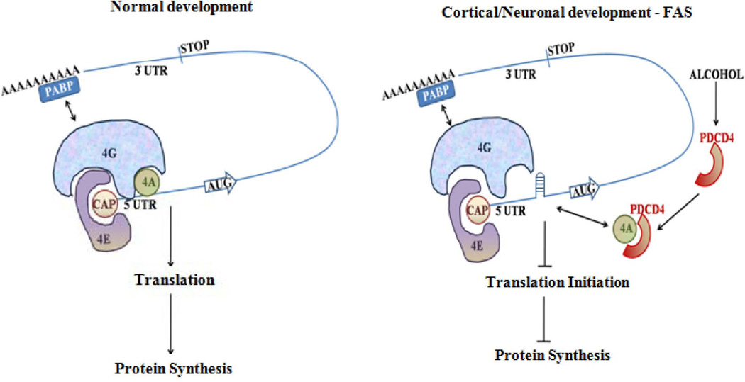
Background: Prenatal exposure to ethanol (EtOH) elicits a range of neuro-developmental abnormalities, microcephaly to behavioral deficits. Impaired protein synthesis has been connected to pathogenesis of EtOH-induced brain damage and abnormal neuron development. However, mechanisms underlying these impairments of protein synthesis are not known. In this study, we illustrate the effects of EtOH on programmed cell death protein 4 (PDCD4), a tumor and translation repressor.

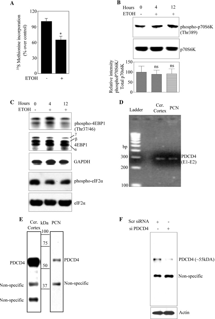
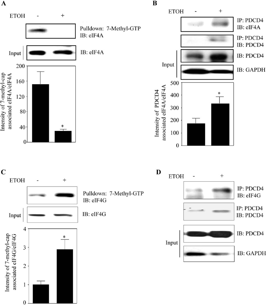
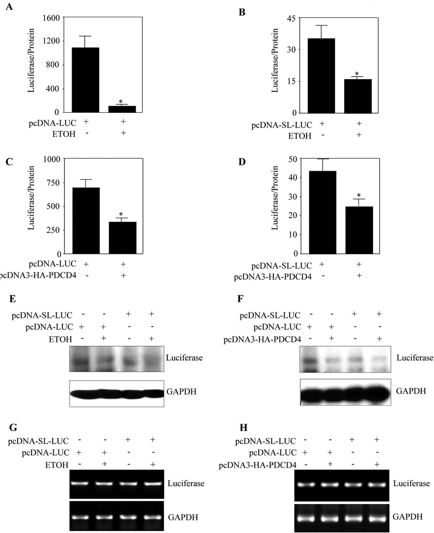
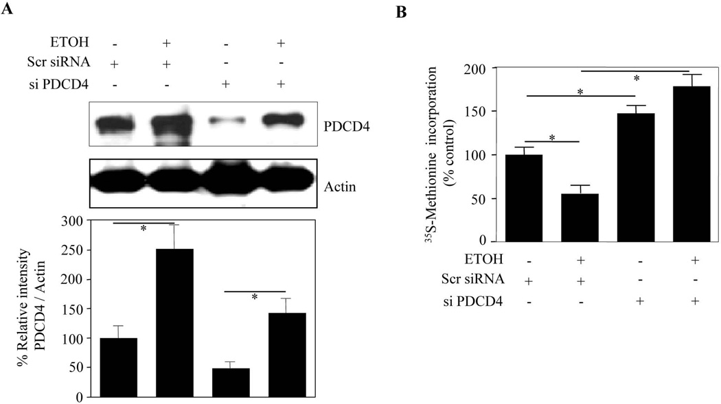
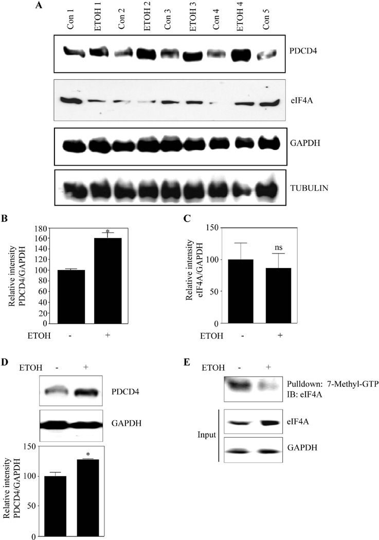
Methods: Primary cortical neurons (PCNs) were treated with 2.5 and 4 mg/ml EtOH for different time points (4 to 24 hours), and PDCD4 expression was detected by Western blotting. Protein synthesis was determined using [(35) S] methionine incorporation assay. Methyl cap pull-down assay was performed to establish the effect of EtOH on association of eukaryotic initiation factor 4A (eIF4A) with capped mRNA. Luciferase assay was performed to determine the in vivo translation. A 2-day acute 5-dose binge model with EtOH (4 g/kg body wt, 25% v/v) was performed in Sprague-Dawley rats at 12-hour intervals and analyzed for PDCD4, eIF4A, and eIF4A-methyl cap association.

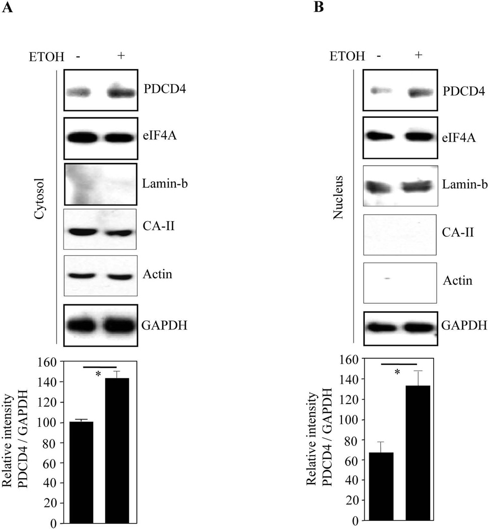
Results: EtOH increased PDCD4 expression in a time- and dose-dependent manner in PCNs, which inhibited the association of eIF4A with methyl cap. EtOH and ectopic PDCD4 expression suppressed in vivo translation in PCNs and RNAi targeting of PDCD4 blocked the inhibitory effect of EtOH on protein synthesis. In utero exposure of pregnant rats to EtOH resulted in a significant increase in PDCD4 in fetal cerebral cortex along with the inhibition of methyl cap-associated eIF4A, compared with isocaloric controls. Increased PDCD4 also occurred in pooled fractions of remaining brain regions.

Conclusions: Our data, for the first time, illustrate that PDCD4 mediates inhibitory effects of EtOH on protein synthesis in PCNs and developing brain.


Figures

Figure 1
Figure 1. Ethanol inhibits protein synthesis in PCNs. Rat brain and fetal cortical neurons express PDCD4
(A) De novo protein synthesis measurement by [35S Methionine] incorporation into proteins in rat PCNs following treatment with ETOH for 24 h. The amount of [35S] radioactivity in the TCA precipitating material was measured and the percentage incorporation of [35S] radioactivity over control was represented. (B) 50µg of protein from untreated and ETOH treated PCN lysates were analyzed using immunoblotting for the extent of phosphorylation in p70S6K at Thr389 using rabbit phospho specific antibody phospho-p70S6K (Thr389) as phosphorylation of Thr389 most closely correlates with p70 kinase activity in vivo. The same blot was stripped and reprobed with total p70S6K which was used as loading control. The bottom panel represents Threonine389 phospho-p70S6K immunoreactivity that was normalized to total p70S6K (p70S6K) and was graphed as percent control. In panel B, ns indicates no significance when compared with untreated control as determined by one way ANOVA. (C) Western analysis was performed using 50µg of protein from untreated and ETOH treated PCN lysates to analyze the extent of phosphorylation in 4EBP1 (Thr37/46) and eIF2α using rabbit phospho specific antibodies. The same blot membrane was stripped and reprobed with corresponding total antibodies that served as loading control. (D) Total RNA isolated from rat brain (RB) and PCN was subjected to one step RT PCR analysis with primers spanning exon1 and exon2 region amplifying a 291 bp product of PDCD4. (E) Western analysis using rabbit anti-PDCD4 polyclonal antibody was performed on total protein lysates from the rat fetal cerebral cortex (75µg) and rat primary fetal cortical neurons (25µg). (F) PCNs were transfected with either non-targeting scrambled siRNA or smart pool mix of four PDCD4 using siPORT amine. Cells were lysed 48 h post transfection and immunoblot analysed with anti-PDCD4 and β-actin.
Figure 2
Figure 2. PDCD4 expression was strongly induced in PCN in response to ETOH
(A) PCNs were treated with ETOH 2.5mg/ml and 4mg/ml for 24 h. PDCD4 protein expression was determined by immunoblot analyses. Immunoblotting with anti-GAPDH was used as loading control. Lower panel depicts densitometric scanning analysis ratio of PDCD4 to GAPDH. Quantification data represents mean ± s.e.m, n=3. (B) PCNs were treated with 4mg/ml ETOH for different time points as indicated. Western analysis for PDCD4 and GAPDH was performed. Lower panel indicate the relative intensity of PDCD4 normalized to GAPDH (mean ± s.e.m, n=3). (C) 5 DIV PCNs were treated with ETOH (4mg/ml) for 24 h and the culture media containing ETOH were replaced with normal culture media for additional 12 h and 24 h. Equal amount of lysates were analyzed for PDCD4 and GAPDH protein expression. One way ANOVA was performed to establish statistical significance. In A, B * - P<0.05, when compared with untreated controls; ns-not significant compared with untreated control.
Figure 3
Figure 3. Ethanol increases cytoplasmic and nuclear content of PDCD4
Western analysis using PDCD4, eIF4A, GAPDH, actin, CA-II and lamin b1 polyclonal antibodies was performed on cytosolic extracts (a) and nuclear extracts (b) from control and ETOH exposed PCNs. In both A & B, the bottom panel depicts the densitometric scanning ratio of PDCD4/ GAPDH intensities. Student’s t-test determined the significance of treatment. * - represents P<0.05 vs untreated controls (mean ± s.e.m, n=3).
Figure 4
Figure 4. ETOH induced PDCD4 sequesters and inhibits eIF4A association with methyl cap mRNA
(A) PCNs treated with or without ETOH for 24 h were used in a pull-down assay with 7-methyl-GTP Sepharose 4B beads which simulates cap mRNA in vitro. 600µg of protein was used to assess the cap-associated eIF4A by immunoblotting. Lower panel depicts the quantification data and Student’s t test was performed to determine the significance of treatment. (B) Methyl cap associated eIF4A depleted supernatants from methyl cap assay were immunoprecipitated with anti-PDCD4 and immunoblotted with antibodies to eIF4A or PDCD4. Equal loading of input samples was confirmed by measuring GAPDH expression. Lower panel represent the densitometric scanning ratio of PDCD4/ GAPDH intensities (mean ± s.e.m, n=6). (C) Methyl cap assay was performed as in Fig. 4A and the cap-associated eIF4G was assessed by immunoblotting. Lower panel is the quantification data of cap-associated eIF4G to direct eIF4G and statistical significance was assessed by Student’s t test. (D) Supernatant from Fig. 4C was immunoprecipitated with anti-PDCD4 and immunoblotted with eIF4G, PDCD4 and GAPDH. In panels A–C, Student’s t test was performed to determine the significance of treatment. * - P<0.05, when compared with untreated controls.
Figure 5
Figure 5. ETOH and transient overexpression of PDCD4 inhibits in vivo translation in PCNs
(A) PCNs were transfected with non-structured mRNA luciferase construct using Fugene HD. 24 h post transfection the cells were treated with or without ETOH (4mg/ml) for additional 24 h. Luciferase activity was measured following normalization to protein levels. (B) PCNs were transfected with structured mRNA luciferase construct using Fugene HD. 24 h following transfection, the cells were treated with or without ETOH (4mg/ml) for additional 24 h. Luciferase activity was determined and the activity was expressed as Luciferase/protein. (C) PCNs were co-transfected with non-stem loop mRNA luciferase construct and PDCD4 expression construct using Fugene HD. 48 h post transfection the cells were processed for determination of luciferase activity. The results were plotted as luciferase/protein values. (D) PCNs were co-transfected with stem-loop mRNA luciferase construct and PDCD4 expression construct using Fugene HD. 48 h following transfection the luciferase activity was determined and the activity was normalized to protein levels. (E) Equal amount of protein samples from 5A, 5B were resolved in SDS-PAGE electrophoresis and immunoblotted for luciferase protein and GAPDH expression. (F) Protein lysates from 5C, 5D were analyzed for luciferase protein and GAPDH expression by immunoblotting. (G) PCNs were transfected and treated as in 5A and 5B. At the end of the experimental period, one step RT-PCR analysis was performed for luciferase mRNA and GAPDH mRNA expression. (H) Total RNA from neurons transfected as in 5C and 5D were subjected for one step RT-PCR analysis with primers specific for luciferase and GAPDH. In Panels A–D, Student’s t test was performed to determine the significance of treatment. * - P<0.05 compared with untreated control (mean ± s.e.m, n=6).
Figure 6
Figure 6. RNAi mediated PDCD4 downregulation reversed ethanol induced suppression of protein translation in PCNs
(A) PCNs were transfected with either non-targeting scramble siRNA or smart pool mix of four siPDCD4 using siPORT amine. 24 h post transfection of scrambled siRNA or siPDCD4 the cells were treated with or without ETOH (4mg/ml) for additional 24 h. Protein extracts were then immunoblot analysed with anti-PDCD4 and anti-actin. (B) PCNs were transfected and treated as in 6A and 2h before the termination of the experiment the cells were labeled with 10 µCi/ml [35S] methionine and processed for [35S Methionine] incorporation into proteins. The amount of [35S] radioactivity in the TCA precipitating material was measured and the percentage incorporation of [35S] radioactivity over control was represented. One way ANOVA was performed to determine the significance of treatment. * - represents P<0.05 (mean ± s.e.m, n=6).
Figure 7
Figure 7. Prenatal ethanol exposure increases PDCD4 expression in fetal brain cortex and rest of the brain
(A) Pregnant rats (Sprague-Dawley) at embryonic day 16 (E16) were administered ETOH (4g/kg body weight) or isocaloric dextrose by gastric intubation at 12 h intervals for two days. At E18 brain cortex from embryos were dissected and processed for PDCD4, eIF4A, GAPDH, Tubulin protein expression by immunoblotting (n=6). (B) This panel illustrates the densitometric scanning ratio of PDCD4/GAPDH intensities. Student’s t test was performed to determine the significance of treatment. * P<0.05 compared with isocaloric dextrose administered animals (mean ± s.e.m, n=6). (C) Densitometric scanning ratio of eIF4A/ GAPDH intensities. Statistical analysis was determined by Student’s t test. ns - represent not significant compared with untreated controls (mean ± s.e.m, n=3). (D) Equal amount of protein lysates from rest of the brain devoid of cortex was separated by SDS-PAGE electrophoresis and immunoblotted with PDCD4 and GAPDH. Lower panel show the densitrometric analysis of PDCD4/GAPDH band intensities. Statistical analysis using Student’s t test indicates * P<0.05 when compared with isocaloric dextrose administered animals. (E) 225µg of cortical brain lysates obtained from 7A is processed for methyl cap analysis as in Fig. 4A and a representative immunoblot is given.
Figure 8
Figure 8. Schematic showing role of PDCD4 in FAS model
During normal development, when PDCD4 expression is maintained at physiological level, eIF4A is available to bind to cap structure and unwinds the mRNA enabling normal translational process. In the event of ETOH abuse during pregnancy, increase in endogenous PDCD4 levels could sequester eIF4A thus hindering the association of eIF4A to the cap structure. Thus the complex structure of mRNA 5’ UTR is left unresolved leading to hampered scanning of ribosomes, inhibiting translation initiation.

Similar articles

Cited by

References

    1. Archibald SL, Fennema-Notestine C, Gamst A, Riley EP, Mattson SN, Jernigan TL. Brain dysmorphology in individuals with severe prenatal alcohol exposure. Dev Med Child Neurol. 2001;43:148–154. - PubMed
    1. Böhm M, Sawicka K, Siebrasse JP, Brehmer-Fastnacht A, Peters R, Klempnauer KH. The transformation suppressor protein Pdcd4 shuttles between nucleus and cytoplasm and binds RNA. Oncogene. 2003;22:4905–4910. - PubMed
    1. Bonner AB, Dalwai S, Marway JS, Preedy VR. Acute exposure to the nutritional toxin alcohol reduces brain protein synthesis in vivo. Metabolism. 2003;52:389–396. - PubMed
    1. Bookstein FL, Streissguth AP, Sampson PD, Connor PD, Barr HM. Corpus callosum shape and neuropsychological deficits in adult males with heavy fetal alcohol exposure. Neuroimage. 2002;15:233–251. - PubMed
    1. Chang JH, Cho YH, Sohn SY, Choi JM, Kim A, Kim YC, Jang SK, Cho Y. Crystal structure of the eIF4A-PDCD4 complex. Proc Natl Acad Sci U S A. 2009;106:3148–3153. - PMC - PubMed

Publication types

MeSH terms