Review
. 2011 Aug;7(3):201-12.
doi: 10.2174/157340311798220502.

Stem cell therapy in heart diseases: a review of selected new perspectives, practical considerations and clinical applications

Affiliations
Review

Stem cell therapy in heart diseases: a review of selected new perspectives, practical considerations and clinical applications

Eltyeb Abdelwahid et al. Curr Cardiol Rev. 2011 Aug.

Abstract

Degeneration of cardiac tissues is considered a major cause of mortality in the western world and is expected to be a greater problem in the forthcoming decades. Cardiac damage is associated with dysfunction and irreversible loss of cardiomyocytes. Stem cell therapy for ischemic heart failure is very promising approach in cardiovascular medicine. Initial trials have indicated the ability of cardiomyocytes to regenerate after myocardial injury. These preliminary trials aim to translate cardiac regeneration strategies into clinical practice. In spite of advances, current therapeutic strategies to ischemic heart failure remain very limited. Moreover, major obstacles still need to be solved before stem cell therapy can be fully applied. This review addresses the current state of research and experimental data regarding embryonic stem cells (ESCs), myoblast transplantation, histological and functional analysis of transplantation of co-cultured myoblasts and mesenchymal stem cells, as well as comparison between mononuclear and mesenchymal stem cells in a model of myocardium infarction. We also discuss how research with stem cell transplantation could translate to improvement of cardiac function.


Figures
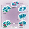
Fig. (1)
Fig. (1)
hESC propagation and in vitro differentiation into CMCs. hESC lines can be propagated continuously in the undifferentiated state when grown on top of an MEF feeder layer. With the Kehat protocol‡ [5], when hESCs are removed from these conditions and grown in suspension, they begin to generate three-dimensional differentiating cell aggregates termed embryoid bodies (EBs). Two weeks after plating on gelatin coated plates, spontaneously contracting areas appear within the EBs. The Mummery protocol* [6], however, uses END-2 cells in the place of MEFs as feeders for hESCs; within 2 weeks, spontaneously contracting areas appear in the hESC-colonies. (hESCs: human Embryonic Stem Cells; MEFs: Mouse Embryo Fibroblasts; EBs: Embryoid Bodies; END2: visceral endoderm-like cells; CMCs: cardiomyocytes)
Fig. (2)
Fig. (2)
Ejection Fraction(EF%) of left ventricle between two groups and in the two periods of evaluation.
Fig. (3)
Fig. (3)
New skeletal fibers (white arrows) and new vessels and endothelial cells (black arrows) identified in a myocardial infarction (MI) (Gomery’s Trichrome, x 200).
Fig. (4)
Fig. (4)
New skeletal fibers(white arrows) identified in an injured myocardium (IM) of Chagas disease. (H&E, X200).
Fig. (5)
Fig. (5)
Angiogenesis: Multiple new vessels after MoSC therapy in the myocardial scar stained by Gomori's Trichrome (x 200).
Fig. (6)
Fig. (6)
New vessel after MeSC in the myocardial scar stained by Gomori's Trichrome (x 400).
Fig. (7)
Fig. (7)
Procedural steps of cell transplantation using trans-coronary venous approach. Left: coronary artery visualization in LAO 30 view; middle: administration of the contrast medium via a guiding catheter placed in the coronary sinus in LAO 30 view, note the balloon inflated at the tip of the guiding catheter to slow the contrast medium outflow; right: placement of TransAccess® catheter system in the anterior interventricular vein and injection of the cell suspension via the IntraLume® microcatheter into anterior wall myocardium – the arrow indicates the microcatheter tip.
Fig. (8)
Fig. (8)
Procedural steps of cell transplantation using trans-coronary venous approach. Left: coronary artery visualization in LAO 30 view; middle: administration of the contrast medium via a non-occlusive guiding catheter placed in the coronary sinus; right: placement of TransAccess® catheter system in the anterior interventricular vein and injection of the cell suspension via the IntraLume® microcatheter into the septum – the arrow indicates the microcatheter tip.
Fig. (9)
Fig. (9)
Intravascular ultrasound image obtained from the cardiac venous site. Please note the visibility of the pericardium (big arrows) and the coronary artery parallel to the vein (small arrows), enabling the orientation of TransAccess® catheter system.

Similar articles

Cited by

References

    1. Reubinoff BE, Itsykson P, Turetsky T, et al. Neural progenitors from human embryonic stem cells. Nat Biotechnol. 2001;19:1134–40. - PubMed
    1. Assady S, Maor G, Amit M, Itskovitz-Eldor J, Skorecki KL, Tzukerman M. Insulin production by human embryonic stem cells. Diabetes. 2001;50:1691–7. - PubMed
    1. Beyer EC, Paul DL, Goodenough DA. Connexin43: a protein from rat heart homologous to a gap junction protein from liver. J Cell Biol. 1987;105:2621–9. - PMC - PubMed
    1. Levenberg S, Golub JS, Amit M, Itskovitz-Eldor J, Langer R. Endothelial cells derived from human embryonic stem cells. Proc Natl Acad Sci USA. 2002;99:4391–6. - PMC - PubMed
    1. Kehat I, Khimovich L, Caspi O, et al. Electromechanical integration of cardiomyocytes derived from human embryonic stem cells. Nat Biotechnol. 2004;22:1282–9. - PubMed

MeSH terms