Review
. 2011 Nov;7(4):234-44.
doi: 10.2174/157340311799960672.

Noninvasive diagnosis of chemotherapy related cardiotoxicity

Affiliations
Review

Noninvasive diagnosis of chemotherapy related cardiotoxicity

Hamilton S Gillespie et al. Curr Cardiol Rev. 2011 Nov.

Abstract

Chemotherapeutic agents reduce mortality and can prevent morbidity in a wide range of malignancies. These agents are, however, associated with toxicities of their own, and the treating physician must remain ever vigilant against the risk outgrowing the benefit of therapy. Thus, pre-treatment evaluation and monitoring for toxicity during and following administration is a fundamental tenet of oncologic practice. Among the most insidious and deadly toxicities of antitumor agents is cardiac toxicity, which in some cases may be irreversible. Early detection of cardiotoxicity allows the treating oncologist to redirect therapy or dose modify, taking into account the cost of a reduction in therapy against the potential of further injury to the patient. In these instances, the role of the cardiologist is to assist and advise the oncologist by providing diagnostic and prognostic information regarding developing cardiotoxicity. This review discusses noninvasive diagnostic options to identify and characterize cardiotoxicity and their use in prognosis and guiding therapy. We also review established protocols for cardiac safety monitoring in the treatment of malignancy.


Figures

Fig. (1)
Fig. (1)
Relationship of left-ventricular ejection fraction, as assessed by echocardiography, in patients undergoing high-dose chemotherapy. Those with any elevation in troponin-I above a cutoff of 0.5 ng/mL after any cycle of chemotherapy demonstrated a significant and persistent drop in left ventricular ejection fraction after roughly 3 months, an effect which persisted out to 7 months. (adapted with permission from Cardinale D et al. JACC 2000; 36(2):517-522)
Fig. (2)
Fig. (2)
Noninvasive assessment of cardiac structure and function. Multiple-gated acquisition (MUGA) images of the right and left ventricles (A). Single photon emission computed tomography (SPECT) assessment of the left ventricle (B). Echocardiography 4-chamber view of the right and left ventricles as obtained from an apical window (C). Cardiac magnetic resonance (CMR) images of the right and left ventricles obtaining utilizing a 1.5 Tesla MRI scanner (D). Coronary CT angiography images, including a volume-rendered image (E), multiplanar reconstruction of the right and left ventricles (F), and maximum intensity projections of the right coronary artery (G) and left anterior descending coronary artery (H).
Fig. (3)
Fig. (3)
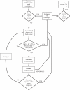
Recommended algorithm for serial monitoring of cardiac function during chemotherapy utilizing cardiovascular imaging techniques. The methodology used to assess LV function is chosen according to the algorithm outlined in Fig. (4). Once a particular modality is chosen, in general, that same method should be used to assess LV function on subsequent cycles.
Fig. (4)
Fig. (4)
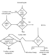
Algortihm for choosing a modality for assessment of left ventricular function. Echocardiography is recommended as the method of choice. If poor image quality limits interpretation, or a fall in EF is detected (see text), then transition to cardiac MRI is recommended. If cardiac MRI is not possible or contraindicated, use of MUGA is indicated. Once a methodology is chosen, we recommend utilizing that methodology for serial assessment. If a fall in EF is detected, transition to the use of cardiac MRI or MUGA to confirm the change is warranted, and if sustained, serial examination with the new modality would be continued. *Aside from availability of appropriate equipment, staff and technicians, contraindications to cardiac MRI include ferromagnetic implants (including certain types of intracranial aneurysm clips), cardiac pacemakers and defibrillators, invasive hemodynamic monitoring devices, certain implanted contraceptive devices, cochlear implants, ferromagnetic shrapnel or other foreign bodies. In the case of gadolinium based contrast administration, contraindications include hepatorenal syndrome, prior liver transplant, and creatinine clearance less than 30 mL/min

Similar articles

Cited by

References

    1. Cardinale D, Colombo A, Sandri MT, et al. Prevention of high-dose chemotherapy-induced cardiotoxicity in high-risk patients by angiotensin-converting enzyme inhibition. Circulation. 2006;114(23):2474–81. - PubMed
    1. Bosch X, Esteve J, Sitges M, et al. Prevention of chemotherapy-induced left ventricular dysfunction with enalapril and carvedilol rationale and design of the OVERCOME trial. J Card Fail. 2011;17(8):643–8. - PubMed
    1. Seidman A, Hudis C, Pierri MK, et al. Cardiac Dysfunction in the Trastuzumab Clinical Trials Experience. Journal of Clinical Oncology. 2002;20(5):1215–21. - PubMed
    1. Shapira J, Gotfried M, Lishner M, et al. Reduced cardiotoxicity of doxorubicin by a 6-hour infusion regimen. A prospective randomized evaluation. Cancer. 1990;65(4):870–3. - PubMed
    1. Legha SS, Benjamin RS, Mackay B, et al. Reduction of doxorubicin cardiotoxicity by prolonged continuous intravenous infusion. Ann Intern Med. 1982; 96(2 ):133–9. - PubMed

MeSH terms