

Altered expression and splicing of Ca(2+) metabolism genes in myotonic dystrophies DM1 and DM2
- PMID: 22758909
- PMCID: PMC3882430
- DOI: 10.1111/j.1365-2990.2012.01289.x
Altered expression and splicing of Ca(2+) metabolism genes in myotonic dystrophies DM1 and DM2
Abstract
Aims: Myotonic dystrophy types 1 and 2 (DM1 and DM2) are multisystem disorders caused by similar repeat expansion mutations, with similar yet distinct clinical features. Aberrant splicing of multiple effector genes, as well as dysregulation of transcription and translation, has been suggested to underlie different aspects of the complex phenotypes in DM1 and DM2. Ca(2+) plays a central role in both muscle contraction and control of gene expression, and recent expression profiling studies have indicated major perturbations of the Ca(2+) signalling pathways in DM. Here we have further investigated the expression of genes and proteins involved in Ca(2+) metabolism in DM patients, including Ca(2+) channels and Ca(2+) binding proteins.
Methods: We used patient muscle biopsies to analyse mRNA expression and splicing of genes by microarray expression profiling and RT-PCR. We studied protein expression by immunohistochemistry and immunoblotting.
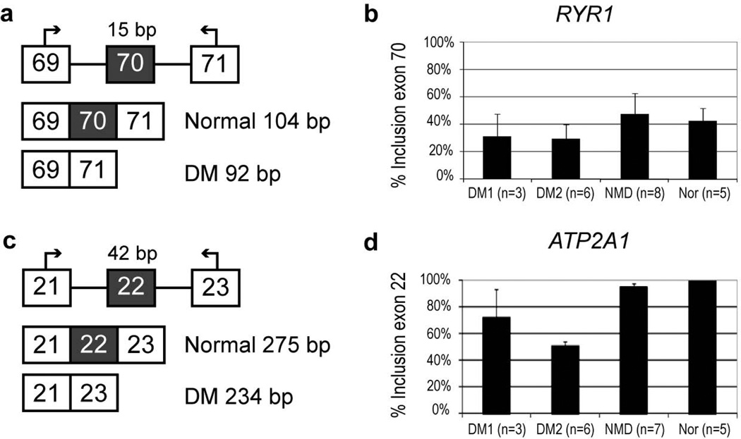
Results: Most of the genes studied showed mRNA up-regulation in expression profiling. When analysed by immunohistochemistry the Ca(2+) release channel ryanodine receptor was reduced in DM1 and DM2, as was calsequestrin 2, a sarcoplasmic reticulum lumen Ca(2+) storage protein. Abnormal splicing of ATP2A1 was more pronounced in DM2 than DM1.
Conclusions: We observed abnormal mRNA and protein expression in DM affecting several proteins involved in Ca(2+) metabolism, with some differences between DM1 and DM2. Our protein expression studies are suggestive of a post-transcriptional defect(s) in the myotonic dystrophies.
© 2012 The Authors. Neuropathology and Applied Neurobiology © 2012 British Neuropathological Society.
Conflict of interest statement
Figures


References
-
- Harper PS. Myotonic Dystrophy. 3rd edn. London: Saunders; 2001.
-
- Udd B, Meola G, Krahe R, Thornton C, Ranum L, Day J, Bassez G, Ricker K. Report of the 115th ENMC workshop: DM2/PROMM and other myotonic dystrophies. 3rd workshop, 14–16 February 2003, Naarden, the Netherlands. Neuromuscul Disord. 2003;13:589–596. - PubMed
-
- Brook JD, McCurrach ME, Harley HG, Buckler AJ, Church D, Aburatani H, Hunter K, Stanton VP, Thirion JP, Hudson T. Molecular basis of myotonic dystrophy: Expansion of a trinucleotide (CTG) repeat at the 3' end of a transcript encoding a protein kinase family member. Cell. 1992;68:799–808. - PubMed
-
- Fu YH, Pizzuti A, Fenwick RG, Jr, King J, Rajnarayan S, Dunne PW, Dubel J, Nasser GA, Ashizawa T, de Jong P. An unstable triplet repeat in a gene related to myotonic muscular dystrophy. Science. 1992;255:1256–1258. - PubMed
-
- Mahadevan M, Tsilfidis C, Sabourin L, Shutler G, Amemiya C, Jansen G, Neville C, Narang M, Barcelo J, O'Hoy K. Myotonic dystrophy mutation: An unstable CTG repeat in the 3' untranslated region of the gene. Science. 1992;255:1253–1255. - PubMed
Publication types
MeSH terms
Substances
Grants and funding
LinkOut - more resources
Full Text Sources
Other Literature Sources
Molecular Biology Databases
Miscellaneous
