. 2012 Oct;13(10):1378-92.
doi: 10.1111/j.1600-0854.2012.01393.x. Epub 2012 Jul 25.

An inventory of peroxisomal proteins and pathways in Drosophila melanogaster

Affiliations

An inventory of peroxisomal proteins and pathways in Drosophila melanogaster

Joseph E Faust et al. Traffic. 2012 Oct.

Abstract

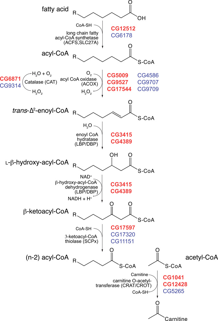
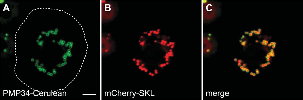
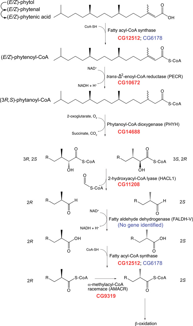
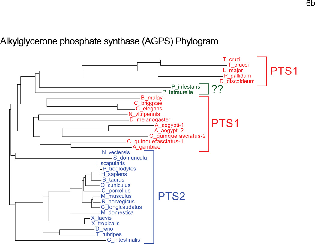
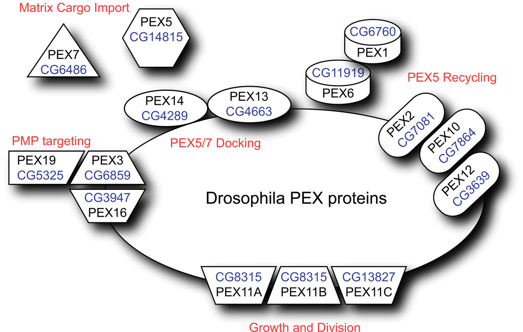
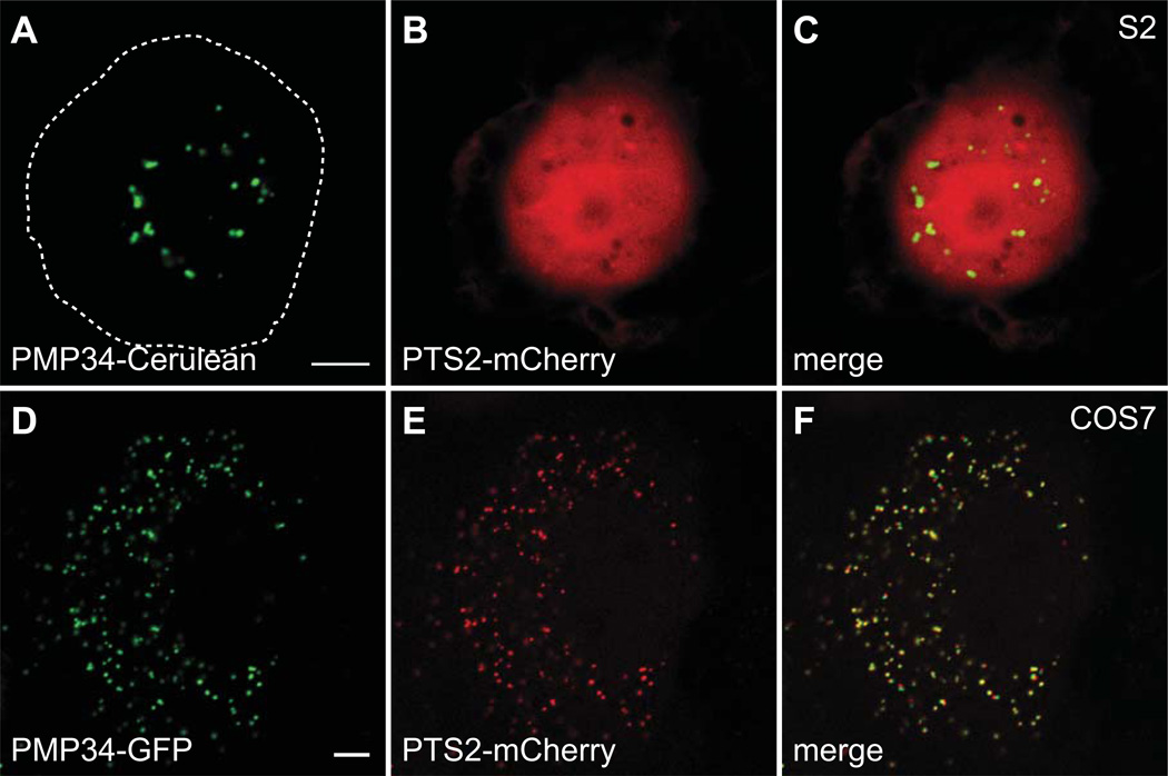
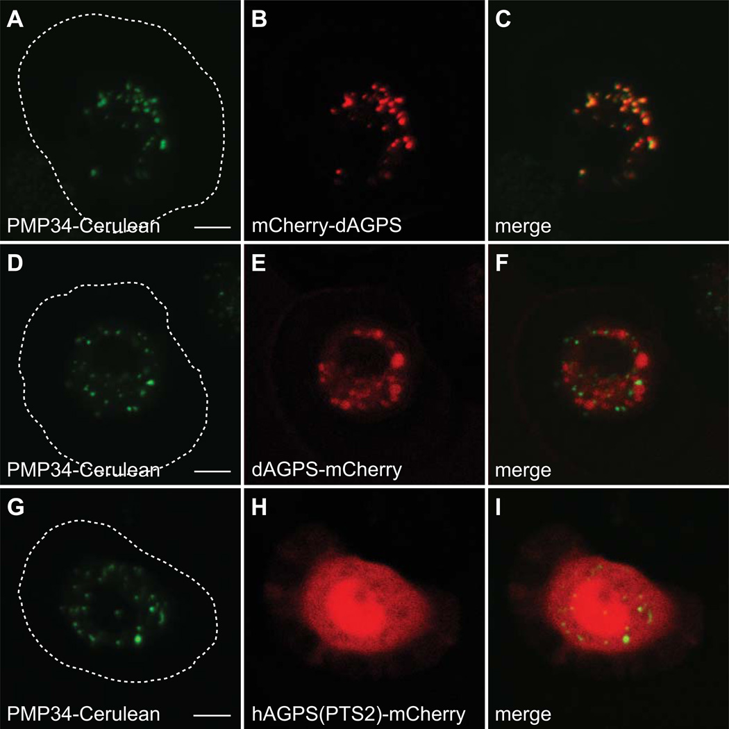
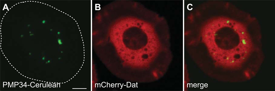
Peroxisomes are ubiquitous organelles housing a variety of essential biochemical pathways. Peroxisome dysfunction causes a spectrum of human diseases known as peroxisome biogenesis disorders (PBD). Although much is known regarding the mechanism of peroxisome biogenesis, it is still unclear how peroxisome dysfunction leads to the disease state. Several recent studies have shown that mutations in Drosophila peroxin genes cause phenotypes similar to those seen in humans with PBDs suggesting that Drosophila might be a useful system to model PBDs. We have analyzed the proteome of Drosophila to identify the proteins involved in peroxisomal biogenesis and homeostasis as well as metabolic enzymes that function within the organelle. The subcellular localization of five of these predicted peroxisomal proteins was confirmed. Similar to Caenorhabditis elegans, Drosophila appears to only utilize the peroxisome targeting signal type 1 system for matrix protein import. This work will further our understanding of peroxisomes in Drosophila and add to the usefulness of this emerging model system.


Figures

Figure 1
Figure 1. Peroxisomal β-oxidation
The steps of peroxisomal β-oxidation of fatty acids are illustrated with the CG numbers for each Drosophila protein listed at each step. The CG numbers are listed in descending order with the best hit(s) in bolded, red text. The percent identities of the Drosophila and human homologs are listed below. ACFS2/SLC27A2/SLC27A4: 22–36%, CAT: 57–65%, ACOX1/2/3: 43–22%, LBP/DBP: 27–53%, SCPx: 35–65%, CRAT/CROT: 25–34%.
Figure 2
Figure 2. mCherry-SKL and PMP34-Cerulean localize to peroxisomes
Plasmids encoding mCherry-SKL and PMP34-Cerulean, under control of the actin 5c promoter, were cotransfected into S2 cells and imaged live by confocal microscopy. (A–C) The mCherry signal is present in a punctate pattern indicating peroxisomal matrix localization. The Cerulean signal is also punctate, but brighter at the edges of the puncta, indicating localization to the peroxisomal membrane. The approximate cell boundary is highlighted with a dashed white line. Scale bar equals 5 µM.
Figure 3
Figure 3. An mCherry-Acyl-CoA Oxidase (CG17544) Fusion Protein localizes to peroxisomes in S2 Cells
Plasmids encoding mCherry-CG17544 and PMP34-Cerulean, under control of the actin 5c promoter, were cotransfected into S2 cells and imaged live by confocal microscopy. (A–C) Colocalizaion of mCherry and Cerulean indicates that mCherry-CG17544 localizes to peroxisomes. The approximate cell boundary is highlighted with a dashed white line. Scale bar equals 5 µM.
Figure 4
Figure 4. An mCherry-Bifunctional Protein (CG4389) Fusion Protein localizes to peroxisomes in S2 Cells
Plasmids encoding mCherry-CG4389 and PMP34-Cerulean, under control of the actin 5c promoter, were cotransfected into S2 cells and imaged live by confocal microscopy. (A–C) Colocalization of mCherry and Cerulean indicates that mCherry-CG4389 localizes to peroxisomes. The approximate cell boundary is highlighted with a dashed white line. Scale bar equals 5 µM.
Figure 5
Figure 5. Peroxisomal α-oxidation Pathway
The steps of peroxisomal α-oxidation are illustrated with the CG numbers for each Drosophila protein listed at each step. The CG numbers are listed in descending order with the best hit(s) in bolded, red text. The percent identities of the Drosophila and human homologs are listed below. ACFS2/SLC27A2/4: 22–36%, PECR: 27%, PHYH: 13%, HACL1: 53%, AMACR: 49%.
Figure 6
Figure 6. Peroxisomal ether lipid synthesis Pathway
A. The steps of peroxisomal ether lipid synthesis are illustrated with the CG numbers for each Drosophila protein listed at the right of each step. The CG numbers are listed in descending order with the best hit(s) in bolded, red text. No homolog of 1-alkyldihydroxyacetone 3-phosphate reductase could be identified in the Drosophila genome. B. A Phylogram of alkylglycerone phosphate synthase (AGPS) from multiple species was generated with ClustalW2. All vertebrate AGPS sequences analyzed contain a PTS2, while most non-vertebrate AGPS sequences, including Drosophila, have a PTS1. P. tetraurelia and P. infestans do not have identifiable PTS motifs. The percent identities of the Drosophila and human homologs are listed below. GNPAT: 25%, FAR1/2: 25–38%, AGPS: 50%.
Figure 7
Figure 7. Drosophila Peroxins
Proteins involved in peroxisome biogenesis, protein import, and growth and division are illustrated. CG numbers for the Drosophila homologs are shown.
Figure 8
Figure 8. PTS2-mCherry localizes to the cytoplasm in S2 cells and peroxisomes in COS7 cells
Plasmids encoding PTS2-mCherry and PMP34-Cerulean, under control of the actin 5c promoter, were cotransfected into S2 cells and imaged live by confocal microscopy. (A–C) The mCherry signal does not colocalize with Cerulean and is diffuse throughout the cell indicating that PTS2-mCherry localizes to the cytoplasm. Plasmids encoding PTS2-mCherry and PMP34-GFP, under control of the CMV promoter, were cotransfected into COS7 cells and imaged live by confocal microscopy. (D–F) The PTS2-mCherry signal colocalizes with PMP34-GFP, indicating peroxisomal localization. The approximate S2 cell boundary is highlighted with a dashed white line. Scale bar equals 5 µM.
Figure 9
Figure 9. Alkylglycerone phosphate synthase (AGPS) localization in S2 cells
Plasmids encoding mCherry-dAGPS, dAGPS-mCherry, or hAGPS(PTS2)-mCherry, under control of the actin 5c promoter, were transfected into S2 cells stably expressing PMP34-Cerulean and imaged live by confocal microscopy. (A–C) The mCherry-dAGPS signal colocalizes with Cerulean, indicating peroxisomal localization due to the PTS1 (AKL) of dAGPS. (D–F) The dAGPS-mCherry signal does not colocalize with Cerulean, indicating that dAGPS does not sort to peroxisomes via an N-terminal PTS2 motif. (G–I) The hAGPS(PTS2)-mCherry signal does not colocalize with Cerulean, indicating that the PTS2 of hAGPS is not sufficient to target mCherry to peroxisomes in S2 cells. The approximate cell boundary is highlighted with a dashed white line. Scale bar equals 5 µM.
Figure 10
Figure 10. Alkylglycerone phosphate synthase (AGPS) localization in COS7 cells
Plasmids encoding PMP34-GFP and either mCherry-dAGPS, dAGPS-mCherry, or hAGPS(PTS2)-mCherry, under control of the CMV promoter, were cotransfected into COS7 cells and imaged live by confocal microscopy. (A–C) The mCherry-dAGPS signal colocalizes with GFP, indicating peroxisomal localization due to the PTS1 (AKL) of dAGPS. (D–F) The dAGPS-mCherry signal does not colocalize with GFP, indicating that dAGPS does not sort to peroxisomes via an N-terminal PTS2 motif. (G–I) The hAGPS(PTS2)-mCherry signal colocalizes with Cerulean, indicating that the PTS2 of hAGPS is sufficient to target mCherry to peroxisomes in COS7 cells. Scale bar equals 5 µM.
Figure 11
Figure 11. Cu/Zn superoxide dismutase carries a PTS1 motif that partially localizes mCherry to peroxisomes
Plasmids encoding PMP34-Cerulean and either mCherry-Sod-PTS1(wild type), mCherry-Sod-PTS1(AKL), or mCherry-Sod-PTS1(ΔAKV) under the control of the actin 5c promoter were cotransfected into S2 cells and imaged by live confocal microscopy. (A–C) The wild-type PTS1 of Cu/Zn Sod localizes mCherry to both the cytoplasm and peroxisomes. Changing the PTS1 to AKL (D–F), shifts mCherry localization to the peroxisome exclusively. Removing the PTS1 (G–I) results in a cytoplasmic localization of mCherry. The PTS1 of Cu/Zn Sod may be a non-optimal sequence and lead to dual localization of this protein. (J–L) Plasmids encoding mCherry-SKL and GFP-CCS-PTS1 under the control of the actin 5c promoter were cotransfected into S2 cells and imaged by live confocal microscopy. The 10 C-terminal amino acids of Copper chaperone for Sod (CCS, CG17753) fused to mCherry is also dually localized to the cytoplasm and peroxisomes. The approximate cell boundary is highlighted with a dashed white line. Scale bar equals 5 µM.
Figure 12
Figure 12. A mCherry-Dopamine N-acetyltransferase (CG3318) Fusion Protein localizes to the cytoplasm in S2 Cells
Plasmids encoding mCherry-CG3318 and PMP34-Cerulean, under control of the actin 5c promoter, were cotransfected into S2 cells and imaged live by confocal microscopy. (A–C) The mCherry signal does not colocalize with Cerulean and is diffuse throughout the cell indicating that mCherry-CG3318 localizes to the cytoplasm. The approximate cell boundary is highlighted with a dashed white line. Scale bar equals 5 µM.

Similar articles

Cited by

References

    1. Wanders RJ, Waterham HR. Biochemistry of mammalian peroxisomes revisited. Annu Rev Biochem. 2006;75:295–332. - PubMed
    1. Hoepfner D, Schildknegt D, Braakman I, Philippsen P, Tabak HF. Contribution of the endoplasmic reticulum to peroxisome formation. Cell. 2005;122(1):85–95. - PubMed
    1. Platta HW, Erdmann R. Peroxisomal dynamics. Trends Cell Biol. 2007;17(10):474–484. - PubMed
    1. Gould SG, Keller GA, Subramani S. Identification of a peroxisomal targeting signal at the carboxy terminus of firefly luciferase. J Cell Biol. 1987;105(6 Pt 2):2923–2931. - PMC - PubMed
    1. Dammai V, Subramani S. The human peroxisomal targeting signal receptor, Pex5p, is translocated into the peroxisomal matrix and recycled to the cytosol. Cell. 2001;105(2):187–196. - PubMed

Publication types

MeSH terms

LinkOut - more resources