. 2012 Jul 18;23(7):1478-87.
doi: 10.1021/bc300130z. Epub 2012 Jul 3.

M13 bacteriophage display framework that allows sortase-mediated modification of surface-accessible phage proteins

Affiliations

M13 bacteriophage display framework that allows sortase-mediated modification of surface-accessible phage proteins

Gaelen T Hess et al. Bioconjug Chem. .

Abstract

We exploit bacterial sortases to attach a variety of moieties to the capsid proteins of M13 bacteriophage. We show that pIII, pIX, and pVIII can be functionalized with entities ranging from small molecules (e.g., fluorophores, biotin) to correctly folded proteins (e.g., GFP, antibodies, streptavidin) in a site-specific manner, and with yields that surpass those of any reported using phage display technology. A case in point is modification of pVIII. While a phage vector limits the size of the insert into pVIII to a few amino acids, a phagemid system limits the number of copies actually displayed at the surface of M13. Using sortase-based reactions, a 100-fold increase in the efficiency of display of GFP onto pVIII is achieved. Taking advantage of orthogonal sortases, we can simultaneously target two distinct capsid proteins in the same phage particle and maintain excellent specificity of labeling. As demonstrated in this work, this is a simple and effective method for creating a variety of structures, thus expanding the use of M13 for materials science applications and as a biological tool.


Figures
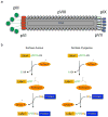
Figure 1
Figure 1. M13 bacteriophage structure and sortase-based reaction schemes
M13 bacteriophage is composed of five capsid proteins. pVIII is the major capsid protein with ~2700 copies in each phage particle. The pVII (light blue) and pIX (blue) are located at one end and start the assembly process, while pIII (green) and pVI (red) are at the other end and cap the phage. Note: the image is not to scale (a). Schematic representation of the mechanism of chemo-enzymatic labeling mediated by Staphylococcus aureus (SrtAaureus-left) or Streptococcus pyogenes (SrtApyogenes-right) (b).
Figure 2
Figure 2. pIII labeling
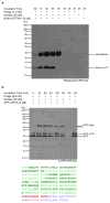
G5-pIII modified phage (there are five copes of pIII/phage particle) was incubated with SrtAaureus and K(biotin)-LPETGG peptide (a), or GFP-LPETG (b), for 3hrs at 37°C or room temperature, respectively. The reactions were monitored by SDS-PAGE under reducing conditions followed by immunoblotting using streptavidin-HRP (a-top panel) or an anti-pIII antibody (a-bottom panel and b). The molecular weight markers are shown on the left. The unidentified anti-pIII reactive protein (*) is most probably a proteolytic fragment of pIII. The identity of the GFP-pIII fusion product was determined by mass-spectrometry. The amino acid sequences of pIII and GFP are shown in blue and green, respectively. The peptides identified are highlighted in bold. The tryptic peptide comprising the GFP C-terminus, followed by the SrtAaureus cleavage site, fused to the N-terminal glycines of pIII is shown in red.
Figure 3
Figure 3. pIX labeling
G5HA-pIX modified phage (there are five copies of pIX/phage particle) was incubated with SrtAaureus and K(biotin)-LPETGG peptide (a), or GFP-LPETG (b), at 37°C and room temperature, respectively, for the times indicated. The reactions were monitored by SDS-PAGE under reducing conditions followed by immunoblotting using streptavidin-HRP (a-top panel) or an anti-HA antibody (a-bottom panel and b). The molecular weight markers are shown on the left. The identity of the GFP-pIX fusion product was determined by mass-spectrometry. The amino acid sequences of pIX and GFP are shown in blue and green, respectively. The peptides identified are highlighted in bold. The AspN digestion-resultant peptide comprising the GFP C-terminus, followed by the SrtAaureus cleavage site, fused to the N-terminal glycines of pIX is shown in red.
Figure 4
Figure 4. pVIII labeling
A2G4-pVIII modified phage (there are 2700 copies of pVIII/phage particle) was incubated with SrtApyogenes and K(biotin)-LPETAA peptide (a), or GFP-LPETA (b), at 37°C for the times indicated in the figure. The reactions were monitored by SDS-PAGE under reducing conditions followed by immunoblotting using streptavidin-HRP (a) or an anti-GFP antibody (b). The molecular weight markers are shown on the left. The unidentified anti-GFP reactive protein (*) is attributed to proteolyzed GFP forming an intermediate with SrtApyogenes. The identity of the GFP-pVIII fusion product was determined by mass-spectrometry. The amino acid sequences of pVIII and GFP are shown in blue and green, respectively. The peptides identified are highlighted in bold. The tryptic peptide comprising the GFP C-terminus, followed by the SrtApyogenes cleavage site, fused to the N-terminal alanines of pVIII is shown in red.
Figure 5
Figure 5. Creation of a multi-phage structure
Schematic representation of the strategy used to build a lampbrush structure (a). Upon labeling of the N-terminus of pIII with streptavidin and of the N-terminus of pVIII with biotin using sortase-mediated reactions, the phage were mixed (see Experimental Procedures section for details). The resulting product was visualized by dynamic light scattering (b) and by atomic force microscopy (c).
Figure 6
Figure 6. Dual labeling of phage using orthogonal SrtApyogenes and SrtAaureus
Schematic representation of the strategy used to couple two different moieties to two different capsid proteins (a). Labeling of pVIII with a K(TAMRA)-LPETAA peptide mediated by SrtApyogenes was followed by labeling of pIII with a single domain antibody directed to Class II MHC as a cell targeting moiety and SrtAaureus (see Experimental Procedures section for details). The final product was analyzed by fluorescent scanning imaging to visualize labeling of pVIII, followed by immunoblotting using an anti-pIII antibody to monitor the efficiency of labeling (b). There are five copies of pIII/phage particle. The asterisks indicate unidentified anti-pIII reactive proteins, which probably correspond to proteolytic fragments of pIII. Binding of the dual labeled phage to lymphocytic Class II MHC+ cells was observed by flow cytometry (c). The Class II MHC+ enriched cell fraction of the lymph nodes of a C57BL/6 mouse was stained for B220 together with the dual labeled phage (phage-TAMRA-VHH7), TAMRA labeled phage (no cell targeting motif, phage-TAMRA), or anti-Class II MHC directly conjugated to TAMRA (TAMRA-VHH7).

Similar articles

Cited by

References

    1. Sidhu SS. Engineering M13 for phage display. Biomol Eng. 2001;18:57–63. - PubMed
    1. Bratkovic T. Progress in phage display: evolution of the technique and its application. Cell Mol Life Sci. 2010;67:749–67. - PMC - PubMed
    1. Burritt JB, Quinn MT, Jutila MA, Bond CW, Jesaitis AJ. Topological mapping of neutrophil cytochrome b epitopes with phage-display libraries. J Biol Chem. 1995;270:16974–80. - PubMed
    1. Barry MA, Dower WJ, Johnston SA. Toward cell-targeting gene therapy vectors: selection of cell-binding peptides from random peptide-presenting phage libraries. Nat Med. 1996;2:299–305. - PubMed
    1. Jaye DL, Nolte FS, Mazzucchelli L, Geigerman C, Akyildiz A, Parkos CA. Use of real-time polymerase chain reaction to identify cell- and tissue-type-selective peptides by phage display. Am J Pathol. 2003;162:1419–29. - PMC - PubMed

Publication types

MeSH terms