Inflammation modulates expression of laminin in the central nervous system following ischemic injury
- PMID: 22759265
- PMCID: PMC3414761
- DOI: 10.1186/1742-2094-9-159
Inflammation modulates expression of laminin in the central nervous system following ischemic injury
Abstract
Background: Ischemic stroke induces neuronal death in the core of the infarct within a few hours and the secondary damage in the surrounding regions over a long period of time. Reduction of inflammation using pharmacological reagents has become a target of research for the treatment of stroke. Cyclooxygenase 2 (COX-2), a marker of inflammation, is induced during stroke and enhances inflammatory reactions through the release of enzymatic products, such as prostaglandin (PG) E2.
Methods: Wild-type (WT) and COX-2 knockout (COX-2KO) mice were subjected to middle cerebral artery occlusion (MCAO). Additionally, brain slices derived from these mice or brain microvascular endothelial cells (BMECs) were exposed to oxygen-glucose deprivation (OGD) conditions. The expression levels of extracellular matrix (ECM) proteins were assessed and correlated with the state of inflammation.
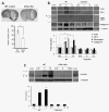
Results: We found that components of the ECM, and specifically laminin, are transiently highly upregulated on endothelial cells after MCAO or OGD. This upregulation is not observed in COX-2KO mice or WT mice treated with COX-2 inhibitor, celecoxib, suggesting that COX-2 is associated with changes in the levels of laminins.
Conclusions: Taken together, we report that transient ECM remodeling takes place early after stroke and suggest that this increase in ECM protein expression may constitute an effort to revascularize and oxygenate the tissue.
Figures


Similar articles
-
MiR-377 Regulates Inflammation and Angiogenesis in Rats After Cerebral Ischemic Injury.J Cell Biochem. 2018 Jan;119(1):327-337. doi: 10.1002/jcb.26181. Epub 2017 Sep 22. J Cell Biochem. 2018. PMID: 28569430
-
Acetylbritannilactone Modulates MicroRNA-155-Mediated Inflammatory Response in Ischemic Cerebral Tissues.Mol Med. 2015 Mar 18;21(1):197-209. doi: 10.2119/molmed.2014.00199. Mol Med. 2015. PMID: 25811992 Free PMC article.
-
NOD2 is involved in the inflammatory response after cerebral ischemia-reperfusion injury and triggers NADPH oxidase 2-derived reactive oxygen species.Int J Biol Sci. 2015 Mar 25;11(5):525-35. doi: 10.7150/ijbs.10927. eCollection 2015. Int J Biol Sci. 2015. PMID: 25892960 Free PMC article.
-
Long Noncoding RNA Malat1 Regulates Cerebrovascular Pathologies in Ischemic Stroke.J Neurosci. 2017 Feb 15;37(7):1797-1806. doi: 10.1523/JNEUROSCI.3389-16.2017. Epub 2017 Jan 16. J Neurosci. 2017. PMID: 28093478 Free PMC article.
-
Cyclooxygenase inhibition in ischemic brain injury.Curr Pharm Des. 2008;14(14):1401-18. doi: 10.2174/138161208784480216. Curr Pharm Des. 2008. PMID: 18537663 Review.
Cited by
-
Basement Membrane Changes in Ischemic Stroke.Stroke. 2020 Apr;51(4):1344-1352. doi: 10.1161/STROKEAHA.120.028928. Epub 2020 Mar 3. Stroke. 2020. PMID: 32122290 Free PMC article. Review. No abstract available.
-
Regulation of the Immune System by Laminins.Trends Immunol. 2017 Nov;38(11):858-871. doi: 10.1016/j.it.2017.06.002. Epub 2017 Jul 3. Trends Immunol. 2017. PMID: 28684207 Free PMC article. Review.
-
Demonstrating a reduced capacity for removal of fluid from cerebral white matter and hypoxia in areas of white matter hyperintensity associated with age and dementia.Acta Neuropathol Commun. 2020 Aug 8;8(1):131. doi: 10.1186/s40478-020-01009-1. Acta Neuropathol Commun. 2020. PMID: 32771063 Free PMC article.
-
Early Rehabilitation Exercise after Stroke Improves Neurological Recovery through Enhancing Angiogenesis in Patients and Cerebral Ischemia Rat Model.Int J Mol Sci. 2022 Sep 10;23(18):10508. doi: 10.3390/ijms231810508. Int J Mol Sci. 2022. PMID: 36142421 Free PMC article.
-
Recombinant Phage Coated 1D Al2O3 Nanostructures for Controlling the Adhesion and Proliferation of Endothelial Cells.Biomed Res Int. 2015;2015:909807. doi: 10.1155/2015/909807. Epub 2015 May 19. Biomed Res Int. 2015. PMID: 26090458 Free PMC article.
References
-
- Herschman H. Prostaglandin synthase 2. Biochim Biophys Acta. 1999;1299:125–140. - PubMed
Publication types
MeSH terms
Substances
Grants and funding
LinkOut - more resources
Full Text Sources
Molecular Biology Databases
Research Materials

