

Cytotoxic CD8+ T cells and CD138+ plasma cells prevail in cerebrospinal fluid in non-paraneoplastic cerebellar ataxia with contactin-associated protein-2 antibodies
- PMID: 22759321
- PMCID: PMC3464990
- DOI: 10.1186/1742-2094-9-160
Cytotoxic CD8+ T cells and CD138+ plasma cells prevail in cerebrospinal fluid in non-paraneoplastic cerebellar ataxia with contactin-associated protein-2 antibodies
Abstract
Objective: The purpose of this paper is to report a patient with otherwise unexplained cerebellar ataxia with serum antibodies against contactin-associated protein-2 (CASPR-2) and provide a detailed description of the composition of cellular infiltrates in the cerebrospinal fluid (CSF) compared to the peripheral blood (PB). CASPR-2 antibodies strongly labeling axons of cerebellar granule neurons have recently been identified in sera from nine patients with otherwise unexplained progressive cerebellar ataxia with mild to severe cerebellar atrophy.
Design: This is a report of a single case.
Methods: The study methods used were neurologic examination, magnetic resonance imaging, fluorodeoxyglucose positron emisson tomography, lumbar puncture and multicolor flow-cytometry.
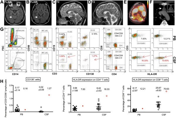
Results: A 23-year-old Caucasian male presented with a two-year history of a progressive cerebellar and brainstem syndrome. Magnetic resonance imaging (MRI) showed pronounced cerebellar atrophy, especially of the medial parts of the hemispheres and the vermis. Cerebral fluorodeoxyglucose positron emission tomography (FDG-PET) showed pronounced hypometabolism of the whole cerebellum. CASPR-2 antibodies were detected in the serum but not the CSF, and none of the staging and laboratory assessments revealed other causes of progressive cerebellar degeneration. Interestingly, flow-cytometry of the CSF as compared to the PB showed increased fractions of CD138+ plasma cells as well as human leukocyte antigen (HLA)-DR+ CD8+ T cells suggesting that both B cells and CD8+ T cells were preferentially recruited to and activated within the CSF- (and putatively central nervous system (CNS)-) compartment.
Conclusion: We confirm the association of CASPR-2 serum antibodies with cerebellar ataxia and provide the first evidence for a combined humoral and cellular immune response in this novel antibody-associated inflammatory CNS disease.
Figures
Similar articles
-
Anti-contactin-associated protein-like 2 antibody-associated cerebellar ataxia: A case report and literature review.J Neuroimmunol. 2021 Apr 15;353:577515. doi: 10.1016/j.jneuroim.2021.577515. Epub 2021 Feb 3. J Neuroimmunol. 2021. PMID: 33640718 Review.
-
[A case of human immunodeficiency virus infection with cerebellar ataxia that suggested by an association with autoimmunity].Rinsho Shinkeigaku. 2016 Apr 28;56(4):255-9. doi: 10.5692/clinicalneurol.cn-000851. Epub 2016 Mar 24. Rinsho Shinkeigaku. 2016. PMID: 27010096 Japanese.
-
Characterization of a Subtype of Autoimmune Encephalitis With Anti-Contactin-Associated Protein-like 2 Antibodies in the Cerebrospinal Fluid, Prominent Limbic Symptoms, and Seizures.JAMA Neurol. 2016 Sep 1;73(9):1115-24. doi: 10.1001/jamaneurol.2016.1585. JAMA Neurol. 2016. PMID: 27428927
-
Paraneoplastic cerebellar syndromes associated with antibodies against Purkinje cells.Int J Neurosci. 2018 Aug;128(8):721-728. doi: 10.1080/00207454.2017.1412967. Epub 2017 Dec 18. Int J Neurosci. 2018. PMID: 29199513
-
Isolated ZIC4 antibodies in paraneoplastic cerebellar syndrome with an underlying ovarian tumor.Arch Neurol. 2011 Aug;68(8):1073. doi: 10.1001/archneurol.2011.176. Arch Neurol. 2011. PMID: 21825246 Review.
Cited by
-
Immune-mediated Cerebellar Ataxias: Practical Guidelines and Therapeutic Challenges.Curr Neuropharmacol. 2019;17(1):33-58. doi: 10.2174/1570159X16666180917105033. Curr Neuropharmacol. 2019. PMID: 30221603 Free PMC article. Review.
-
Promise, Progress, and Pitfalls in the Search for Central Nervous System Biomarkers in Neuroimmunological Diseases: A Role for Cerebrospinal Fluid Immunophenotyping.Semin Pediatr Neurol. 2017 Aug;24(3):229-239. doi: 10.1016/j.spen.2017.08.001. Epub 2017 Aug 12. Semin Pediatr Neurol. 2017. PMID: 29103430 Free PMC article. Review.
-
Role and Relevance of Cerebrospinal Fluid Cells in Diagnostics and Research: State-of-the-Art and Underutilized Opportunities.Diagnostics (Basel). 2021 Dec 30;12(1):79. doi: 10.3390/diagnostics12010079. Diagnostics (Basel). 2021. PMID: 35054246 Free PMC article. Review.
-
Altered Cerebellar Response to Somatosensory Stimuli in the Cntnap2 Mouse Model of Autism.eNeuro. 2021 Oct 21;8(5):ENEURO.0333-21.2021. doi: 10.1523/ENEURO.0333-21.2021. Print 2021 Sep-Oct. eNeuro. 2021. PMID: 34593517 Free PMC article.
-
Expanding spectrum of contactin-associated protein 2 (CASPR2) autoimmunity-syndrome of parkinsonism and ataxia.Neurol Sci. 2018 Mar;39(3):455-460. doi: 10.1007/s10072-017-3222-0. Epub 2017 Dec 20. Neurol Sci. 2018. PMID: 29264691
References
-
- Irani SR, Alexander S, Waters P, Kleopa KA, Pettingill P, Zuliani L, Peles E, Buckley C, Lang B, Vincent A. Antibodies to Kv1 potassium channel-complex proteins leucine-rich, glioma inactivated 1 protein and contactin-associated protein-2 in limbic encephalitis, Morvan’s syndrome and acquired neuromyotonia. Brain. 2010;133:2734–2748. - PMC - PubMed
-
- Melzer N, Meuth SG, Wiendl H. Neuron-directed autoimmunity in the central nervous system: entities, mechanisms, diagnostic clues, and therapeutic options. Curr Opin Neurol. 2012;25:341–348. - PubMed
-
- Melzer N, Harder A, Gross CC, Wolfer J, Stummer W, Niederstadt T, Meuth SG, Marziniak M, Grauer OM, Wiendl H. CD4+ T cells predominate in cerebrospinal fluid and leptomeningeal and parenchymal infiltrates in cerebral amyloid beta-related angiitis. Arch Neurol. 2012. Epub ahead of print. - PubMed
Publication types
MeSH terms
Substances
LinkOut - more resources
Full Text Sources
Research Materials
Miscellaneous
