. 2012 Jul 15;26(14):1558-72.
doi: 10.1101/gad.193516.112. Epub 2012 Jul 3.

ZRANB3 is a structure-specific ATP-dependent endonuclease involved in replication stress response

Affiliations

ZRANB3 is a structure-specific ATP-dependent endonuclease involved in replication stress response

Ria Weston et al. Genes Dev. .

Abstract

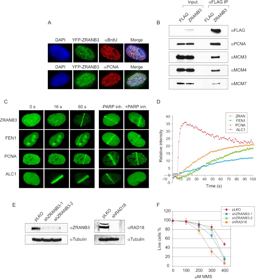
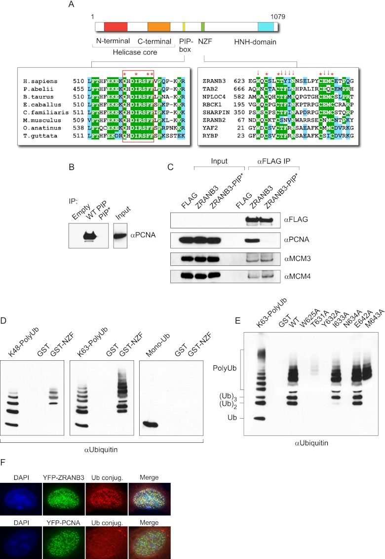
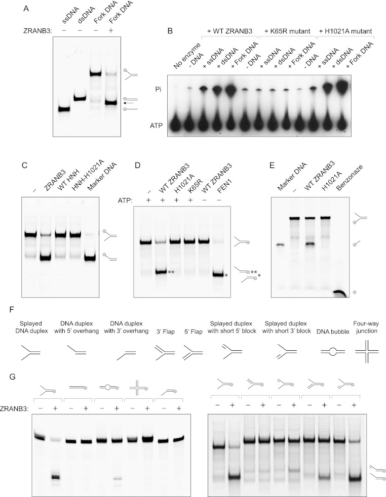
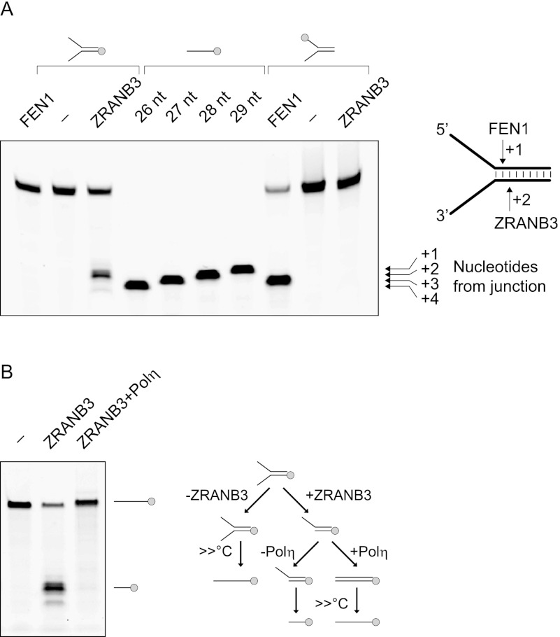
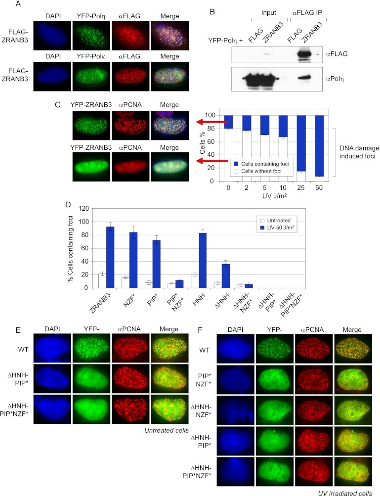
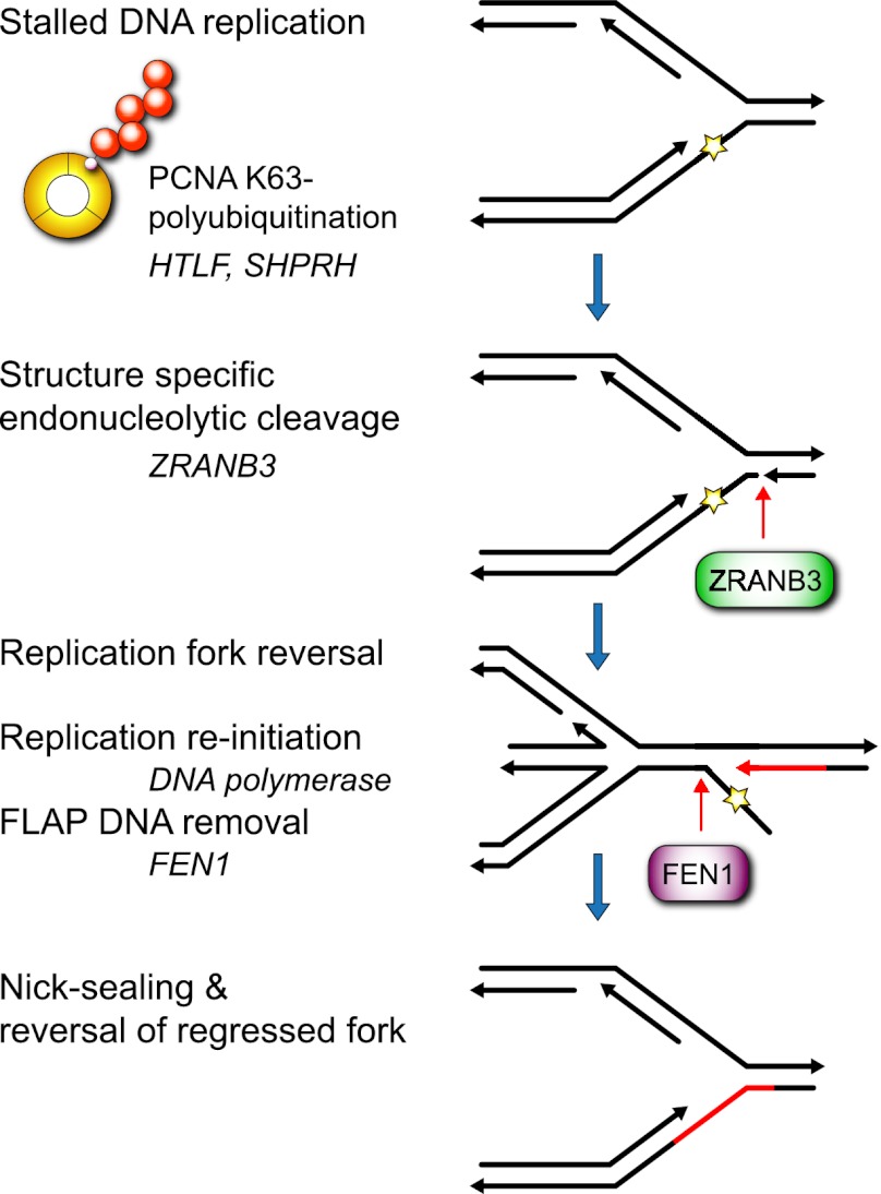
To efficiently duplicate their genomic content, cells must overcome DNA lesions that interfere with processive DNA replication. These lesions may be removed and repaired, rather than just tolerated, to allow continuity of DNA replication on an undamaged DNA template. However, it is unclear how this is achieved at a molecular level. Here we identify a new replication-associated factor, ZRANB3 (zinc finger, RAN-binding domain containing 3), and propose its role in the repair of replication-blocking lesions. ZRANB3 has a unique structure-specific endonuclease activity, which is coupled to ATP hydrolysis. It cleaves branched DNA structures with unusual polarity, generating an accessible 3'-OH group in the template of the leading strand. Furthermore, ZRANB3 localizes to DNA replication sites and interacts with the components of the replication machinery. It is recruited to damaged replication forks via multiple mechanisms, which involve interactions with PCNA, K63-polyubiquitin chains, and branched DNA structures. Collectively, our data support a role for ZRANB3 in the replication stress response and suggest new insights into how DNA repair is coordinated with DNA replication to maintain genome stability.


Figures

Figure 1.
Figure 1.
(A) ZRANB3 targets sites of DNA replication. U2OS cells were transiently transfected with YFP-ZRANB3, pulsed with BrdU for 15 min, and stained with anti-BrdU antibody. Alternatively, cells were transfected with YFP-ZRANB3 and stained against endogenous PCNA. (B) Interaction of ZRANB3 with replication-associated factors. 293T cells were transiently transfected with Flag-tagged ZRANB3 or with empty vector, and extracts were subjected to immunoprecipitation on anti-Flag beads. Immunocomplexes were eluted with 3xFlag peptide and immunoblotted with the indicated antibodies. (C) ZRANB3 is recruited to the sites of laser-induced DNA damage. U2OS cells were transiently transfected with the indicated YFP constructs and analyzed by live-cell imaging. Shown are representative images at the indicated time points post-damage. Recruitment of ZRANB3 is compared with the replication factors PCNA and FEN1 and the SNF2 chromatin remodeling factor ALC1. Cells were also assayed 60 sec after the damage in the presence or absence of a PARP inhibitor. (D) Kinetics of the ZRANB3 recruitment to sites of laser-induced DNA damage, and comparison with the kinetics of PCNA, FEN1 and ALC1. More than 10 cells were analyzed for each construct. (E) Down-regulation of ZRANB3 and RAD18 expression by shRNA. Stable cell lines were created by the expression of empty pLKO-Puro, shZRANB3-1, shZRANB3-2, and shRAD18 vectors. Following selection against puromycin, the expression of ZRANB3 or RAD18 was evaluated by Western blotting. (F) Sensitivity of ZRANB3-deficient cells to MMS.
Figure 2.
Figure 2.
(A) Modular structure of ZRANB3. (Left) Conservation of the PIP-box residues among ZRANB3 proteins is shown in the alignment. (Right) Conservation of the NZF motif includes several representative human proteins. Conserved cysteine residues are marked by asterisks. Arrows indicate mutated residues Trp 625, Thr 631, Tyr 632, Ile 633, Asn 634, Glu 642, and Met 643. (B) Interaction of the ZRANB3 PIP-box motif with PCNA in vitro. Biotinylated PIP-box peptide was bound to streptavidin beads and incubated with recombinant PCNA. The interaction was assayed by Western blotting with PCNA antibody. Mutations of the conserved PIP-box residues (Q519A, F525A, and F526A in PIP*) abrogated the interaction with PCNA. (C) Interaction of ZRANB3 with PCNA is PIP-box-dependent. 293T cells were transiently transfected with empty vector, Flag-tagged wild-type ZRANB3, and the PIP-box mutant (ZRANB3-PIP*). Following immunoprecipitation on anti-Flag beads, immunocomplexes were eluted by 3xFlag peptide and analyzed by Western blotting. Expression of Flag-ZRANB3 is detectable only after immunoprecipitation. (D) Interaction of the NZF motif with polyubiquitin chains in vitro. The wild-type NZF motif was expressed as a GST fusion protein and bound to the GST beads. The beads were then incubated with the monoubiquitin, polyubiquitin K48(2-7), or polyubiquitin K63(2-7) chains. Interactions were assayed by anti-ubiquitin Western blotting. (E) Interactions of mutant NZF motifs with K63–polyubiquitin chains. The experiment was performed as in D. Mutated positions are indicated by arrows in the alignment in A. (F) Colocalization of ZRANB3 with ubiquitin conjugates. U2OS cells were transiently transfected with YFP-ZRANB3 or YFP-PCNA and immunostained with FK2 antibody, which recognizes ubiquitin conjugates but not free ubiquitin.
Figure 3.
Figure 3.
(A) Helicase assay with ZRANB3. Fork DNA (splayed DNA duplex), fluorescently labeled at the 5′ end (as indicated in the picture), was incubated with ZRANB3 in the presence of ATP. After 30 min, an excess of unlabeled complementary ssDNA was added to prevent reannealing of the displaced oligos. Efficient DNA helicase activity is expected to yield fluorescently labeled ssDNA. Labeled ssDNA and dsDNA oligos were used as markers. Products were analyzed by native polyacrylamide gel electrophoresis. The reaction yielded a product of unexpected mobility (indicated by an arrow). (B) ATPase assay with the wild-type ZRANB3, helicase core mutant K65R, and HNH mutant H1021A. Recombinant proteins were incubated with 32P-labeled ATP in the absence or presence of indicated DNA substrates (40 nM). Reaction products were resolved by thin-layer chromatography. (C) Nuclease assay with full-length ZRANB3, isolated wild- type HNH domain, and HNH domain containing the H1021A mutation. Splayed DNA duplex fluorescently labeled at the 5′ end (shown in the picture) was incubated with the indicated proteins in the presence of ATP. Fluorescently labeled DNA duplex with 5′ overhang was used as a marker. Reactions were analyzed by native polyacrylamide gel electrophoresis. (D) Nuclease assay with the full-length ZRANB3 proteins. Wild-type ZRANB3, ATPase dead K65R mutant, and HNH mutant H1021A were incubated with splayed DNA duplex fluorescently labeled at the 5′ end (shown in the picture) in the presence of ATP. Additionally, wild-type ZRANB3 and FEN1 were incubated with the same substrate in the absence of ATP. Reactions were analyzed by native polyacrylamide gel electrophoresis. (E) Nuclease assay with the 3′-end-labeled splayed DNA duplex. Wild-type ZRANB3 was incubated with 3′-end FITC-labeled DNA substrate (as indicated in the picture). As a control, DNA substrate was incubated with benzonase nuclease to yield fluorescently labeled mononucleotides. Shown is a 26-nt marker DNA. (F) Substrates used in nuclease assays with ZRANB3. (G) The ability of ZRANB3 to cleave different DNA substrates. Wild-type ZRANB3 was incubated with the indicated fluorescently labeled DNA substrates in the presence of ATP. Reactions were analyzed by denaturing (left) or native (right) polyacrylamide gel electrophoresis.
Figure 4.
Figure 4.
(A) ZRANB3 cleaves DNA forks in the dsDNA region 2 nt from the branch point. ZRANB3 and FEN1 were incubated with the indicated fluorescently labeled DNA substrates. ATP was added to the reactions with ZRANB3. Reactions were resolved by denaturing polyacrylamide gel electrophoresis, and mobility of the obtained products was compared with the fluorescently labeled ssDNA markers. (B) Cleavage of fork DNA by ZRANB3 yields a 3′-OH end that can be extended by DNA polymerase. The splayed DNA duplex fluorescently labeled at the 5′ end (shown in the picture) was incubated with or without ZRANB3 in the presence of ATP. Polη and dNTPs were then added to the reaction to support extension of the cleaved DNA strand. Products were analyzed by denaturing polyacrylamide gel electrophoresis. Experimental outline is shown on the right.
Figure 5.
Figure 5.
(A) ZRANB3 colocalizes with TLS polymerases Polη and Polκ. U2OS cells were cotransfected with Flag-tagged ZRANB3 and YFP-Polη or YFP-Polκ. Cells were stained with anti-Flag antibody. (B) ZRANB3 immunoprecipitates YFP-Polη from whole-cell extracts. 293T cells were cotransfected with Flag-tagged ZRANB3 and YFP-Polη. Following immunoprecipitation on anti-Flag beads, immunocomplexes were eluted by 3xFlag peptide and analyzed by Western blotting with Polη antibody. (C) ZRANB3 accumulates at replication forks stalled at DNA damage. U2OS cells were transfected with YFP-ZRANB3 and exposed to the indicated doses of UV irradiation. After 6 h, cells were fixed and stained with PCNA antibody. They were analyzed by microscopy, and the percentage of cells containing ZRANB3 foci that colocalized with PCNA was determined. Representative images of cells with and without ZRANB3 foci are shown on the left. (D) Contribution of functional domains within the ZRANB3 structure to its recruitment to sites of DNA replication before and after DNA damage. U2OS cells were transfected with the indicated YFP-ZRANB3 constructs and either untreated or exposed to UV irradiation. After 6 h, cells were fixed and stained with PCNA antibody. The percentages of cells containing ZRANB3 foci that colocalized with PCNA were determined as in C. (E) Representative images of mutant ZRANB3 proteins that do not colocalize with PCNA in the absence of exogenous DNA damage. U2OS cells were transfected with the indicated YFP-ZRANB3 constructs. Cells were stained with PCNA antibody. Merge represents green and red channels. (F) Representative images of mutant ZRANB3 proteins that do not colocalize with PCNA following UV irradiation.
Figure 6.
Figure 6.
Proposed model by which ZRANB3 facilitates replication-associated DNA repair. Separation of DNA strands by replicative helicase exposes the DNA lesion in the leading strand, which blocks processive DNA replication. Unavailability of the complementary DNA strand prevents repair of the exposed lesions by excision repair machinery at this point. E3 ubiquitin ligases HLTF and SHPRH catalyze PCNA K63–polyubiquitination of stalled replication forks, which recruits ZRANB3. At stalled replication forks, ZRANB3 acts as a structure-specific endonuclease and induces a DNA break in the double-stranded region of the replication fork 2 nt from the branching point. This is coordinated with the replication fork regression to prevent its disintegration. Cleavage by ZRANB3 exposes a free 3′-OH group, which can be extended by DNA polymerase to remove the replication-blocking DNA lesion. This leads to the formation of the 5′ overhanging DNA flap, which can be processed by the activity of FEN1. Following nick sealing and reversal of regressed forks, DNA replication resumes on the undamaged DNA template.

Comment in

Similar articles

Cited by

References

    1. Ahel D, Horejsi Z, Wiechens N, Polo SE, Garcia-Wilson E, Ahel I, Flynn H, Skehel M, West SC, Jackson SP, et al. 2009. Poly(ADP-ribose)-dependent regulation of DNA repair by the chromatin remodeling enzyme ALC1. Science 325: 1240–1243 - PMC - PubMed
    1. Alam SL, Sun J, Payne M, Welch BD, Blake BK, Davis DR, Meyer HH, Emr SD, Sundquist WI 2004. Ubiquitin interactions of NZF zinc fingers. EMBO J 23: 1411–1421 - PMC - PubMed
    1. Atkinson J, McGlynn P 2009. Replication fork reversal and the maintenance of genome stability. Nucleic Acids Res 37: 3475–3492 - PMC - PubMed
    1. Bansbach CE, Betous R, Lovejoy CA, Glick GG, Cortez D 2009. The annealing helicase SMARCAL1 maintains genome integrity at stalled replication forks. Genes Dev 23: 2405–2414 - PMC - PubMed
    1. Betous R, Mason AC, Rambo RP, Bansbach CE, Badu-Nkansah A, Sirbu BM, Eichman BF, Cortez D 2012. SMARCAL1 catalyzes fork regression and Holliday junction migration to maintain genome stability during DNA replication. Genes Dev 26: 151–162 - PMC - PubMed

Publication types

MeSH terms