

Phenotype-genotype correlations in mouse models of amelogenesis imperfecta caused by Amelx and Enam mutations
- PMID: 22759786
- PMCID: PMC3718574
- DOI: 10.1159/000336440
Phenotype-genotype correlations in mouse models of amelogenesis imperfecta caused by Amelx and Enam mutations
Abstract
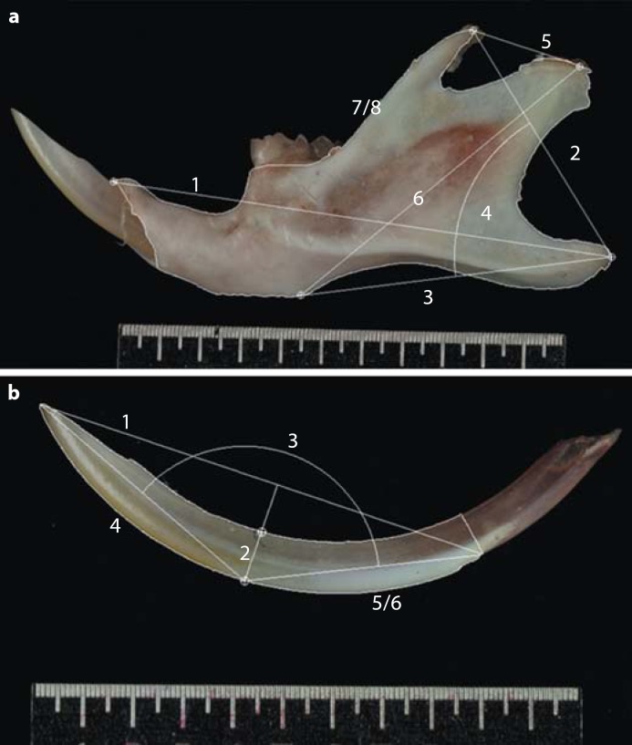
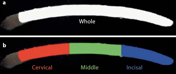
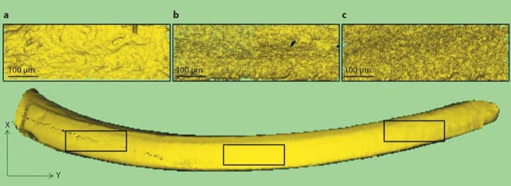
Mutations in human and in mouse orthologous genes Amelx and Enam result in a diverse range of enamel defects. In this study we aimed to investigate the phenotype-genotype correlation between the mutants and the wild-type controls in mouse models of amelogenesis imperfecta using novel measurement approaches. Ten hemi-mandibles and incisors were dissected from each group of Amelx(WT), Amelx(X/Y64H), Amelx(Y/Y64H), Amelx(Y64H/Y64H), and Enam(WT), Enam(Rgsc395) heterozygous and Enam(Rgsc395) homozygous mice. Their macro-morphology, colour and micro-topography were assessed using bespoke 2D and 3D image analysis systems and customized colour and whiteness algorithms. The novel methods identified significant differences (p ≤ 0.05) between the Amelx groups for mandible and incisor size and enamel colour and between the Enam groups for incisor size and enamel colour. The Amelx(WT) mice had the largest mandibles and incisors, followed in descending order of size by the Amelx(X/Y64H), Amelx(Y/Y64H) and Amelx(Y64H/Y64H) mice. Within the Enam groups the Enam(WT) incisors were largest and the Enam(Rgsc395) heterozygous mice were smallest. The effect on tooth morphology was also reflected by the severity of the enamel defects in the colour and whiteness assessment. Amelogenin affected mandible morphology and incisor enamel formation, while enamelin only affected incisors, supporting the multifunctional role of amelogenin. The enamelin mutation was associated with earlier forming enamel defects. The study supported the critical involvement of amelogenin and enamelin in enamel mineralization.
Copyright © 2012 S. Karger AG, Basel.
Figures

Similar articles
-
Enamelin (Enam) is essential for amelogenesis: ENU-induced mouse mutants as models for different clinical subtypes of human amelogenesis imperfecta (AI).Hum Mol Genet. 2005 Mar 1;14(5):575-83. doi: 10.1093/hmg/ddi054. Epub 2005 Jan 13. Hum Mol Genet. 2005. PMID: 15649948
-
Splicing mutations in AMELX and ENAM cause amelogenesis imperfecta.BMC Oral Health. 2023 Nov 20;23(1):893. doi: 10.1186/s12903-023-03508-8. BMC Oral Health. 2023. PMID: 37985977 Free PMC article.
-
A mutation in the mouse Amelx tri-tyrosyl domain results in impaired secretion of amelogenin and phenocopies human X-linked amelogenesis imperfecta.Hum Mol Genet. 2010 Apr 1;19(7):1230-47. doi: 10.1093/hmg/ddq001. Epub 2010 Jan 12. Hum Mol Genet. 2010. PMID: 20067920 Free PMC article.
-
[Alteración del gen AMELX en amelogénesis imperfecta. Una breve revisión].Gac Med Mex. 2019;155(1):101-107. doi: 10.24875/GMM.18003604. Gac Med Mex. 2019. PMID: 30799455 Review. Spanish.
-
Enamelin and autosomal-dominant amelogenesis imperfecta.Crit Rev Oral Biol Med. 2003;14(6):387-98. doi: 10.1177/154411130301400602. Crit Rev Oral Biol Med. 2003. PMID: 14656895 Review.
Cited by
-
Tracking endogenous amelogenin and ameloblastin in vivo.PLoS One. 2014 Jun 16;9(6):e99626. doi: 10.1371/journal.pone.0099626. eCollection 2014. PLoS One. 2014. PMID: 24933156 Free PMC article.
-
Analysis of enamel development using murine model systems: approaches and limitations.Front Physiol. 2014 Sep 17;5:313. doi: 10.3389/fphys.2014.00313. eCollection 2014. Front Physiol. 2014. PMID: 25278900 Free PMC article. Review.
-
Mesenchymal Bmp7 Controls Onset of Tooth Mineralization: A Novel Way to Regulate Molar Cusp Shape.Front Physiol. 2020 Jul 3;11:698. doi: 10.3389/fphys.2020.00698. eCollection 2020. Front Physiol. 2020. PMID: 32719613 Free PMC article.
References
-
- Aldred M.J., Crawford P.J., Roberts E., Thomas N.S. Identification of a nonsense mutation in the amelogenin gene (AMELX) in a family with X-linked amelogenesis imperfecta (AIH1) Hum Gen. 1992;90:413–416. - PubMed
-
- Atchley W.R., Hall B.K. A model for development and evolution of complex morphological structures. Bio Rev. 1991;66:101–157. - PubMed
-
- Barron M.J., Brookes S.J., Kirkham J., Shore R.C., Hunt C., Mironov A., Kingswell N.J., Maycock J., Shuttleworth C.A., Dixon M.J. A mutation in the mouse Amelx tri-tyrosyl domain results in impaired secretion of amelogenin and phenocopies human X-linked amelogenesis imperfecta. Hum Mol Genet. 2010;19:1230–1247. - PMC - PubMed
-
- Bland J.M., Altman D.G. Statistical methods for assessing agreement between two methods of clinical measurement. Lancet. 1986;1:307–310. - PubMed
-
- Bland J.M., Altman D.G. Measuring agreement in method comparison studies. Stat Methods Med Res. 1999;8:135–160. - PubMed
Publication types
MeSH terms
Substances
Grants and funding
LinkOut - more resources
Full Text Sources
Other Literature Sources
Molecular Biology Databases
