

Combining structural brain changes improves the prediction of Alzheimer's disease and mild cognitive impairment
- PMID: 22759808
- PMCID: PMC3490129
- DOI: 10.1159/000339364
Combining structural brain changes improves the prediction of Alzheimer's disease and mild cognitive impairment
Abstract
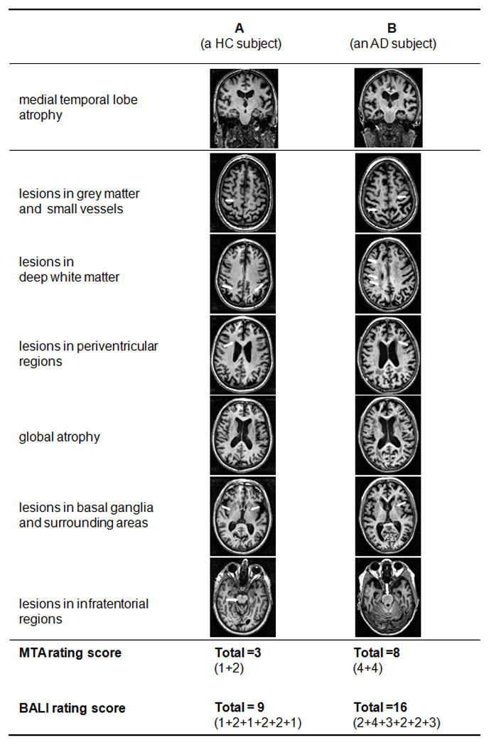
Background: Several structural brain changes have been associated with Alzheimer's disease (AD). This study investigated the prediction of AD by combining multiple brain changes with the hallmark medial temporal lobe atrophy (MTA).
Methods: High-resolution magnetic resonance imaging (MRI) data of people with mild AD (n = 39), mild cognitive impairment (MCI; n = 82), and of healthy controls (HC; n = 58) were obtained at baseline. Among these people, 26 AD, 53 MCI, and 46 HC subjects had 24-month follow-up MRI scans. Bilateral MTA was evaluated using a medial temporal lobe atrophy scale (MTAS). Common changes in the aging brain were summarized using a brain atrophy and lesion index (BALI). The performance of the MTAS, BALI, and a score combining both, using a logistic regression model, were assessed.
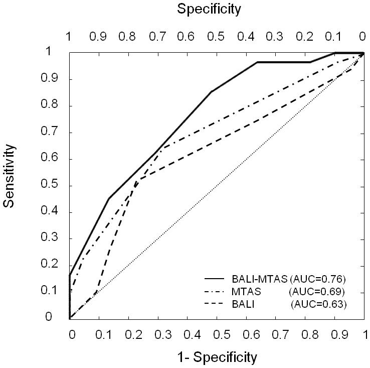
Results: The MTAS and BALI scores were closely correlated (r(2) > 0.56); each differed between the diagnostic groups. Having an unfavorable MTAS score was associated with an increased risk of MCI-AD conversion (OR = 3.71, p = 0.039), adjusted for age, sex, and education; having an unfavorable BALI score marginally contributed to such risks (OR = 4.08, p = 0.080). Combining MTAS and BALI components resulted in a greater OR (8.99, p = 0.007) and an improved predictive accuracy (75.9%, p = 0.002).
Conclusion: Multiple structural changes have an additive effect on AD. The data support potential future roles of combining multiple coexisting structural changes to benefit AD diagnosis, progression monitoring, and/or treatment effect evaluation.
Copyright © 2012 S. Karger AG, Basel.
Figures

References
-
- Dubois B, Feldman HH, Jacova C, Dekosky ST, Barberger-Gateau P, Cummings J, Delacourte A, Galasko D, Gauthier S, Jicha G, Meguro K, O’brien J, Pasquier F, Robert P, Rossor M, Salloway S, Stern Y, Visser PJ, Scheltens P. Research criteria for the diagnosis of Alzheimer’s disease: revising the NINCDS-ADRDA criteria. Lancet Neurol. 2007;6:734–746. - PubMed
-
- Galluzzi S, Talassi E, Belussi M, Scheltens P, van de Pol L, Nobili F, Rodriguez G, Froelich L, Damian M, Martinez-Lage P, Gomez-Isla T, Reynish E, Ousset PJ, Vellas B, Frisoni GB. Multi-center comparison of medial temporal atrophy in patients with Alzheimer’s disease--data from the ICTUS study. Dement Geriatr Cogn Disord. 2008;26:314–322. - PubMed
-
- Korf ES, Wahlund LO, Visser PJ, Scheltens P. Medial temporal lobe atrophy on MRI predicts dementia in patients with mild cognitive impairment. Neurology. 2004;63:94–100. - PubMed
Publication types
MeSH terms
Grants and funding
LinkOut - more resources
Full Text Sources
Medical
