. 2012 Oct;20(10):1953-67.
doi: 10.1038/mt.2012.117. Epub 2012 Jul 3.

Valproic acid confers functional pluripotency to human amniotic fluid stem cells in a transgene-free approach

Affiliations

Valproic acid confers functional pluripotency to human amniotic fluid stem cells in a transgene-free approach

Dafni Moschidou et al. Mol Ther. 2012 Oct.

Expression of concern in

Abstract

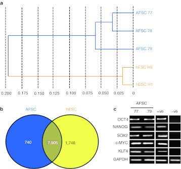
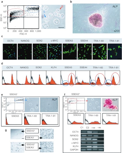
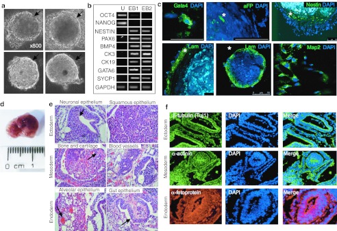
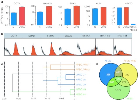
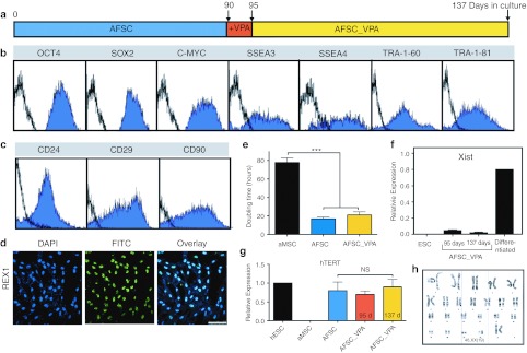
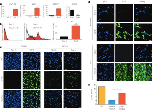
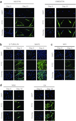
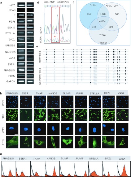
Induced pluripotent stem cells (iPSCs) with potential for therapeutic applications can be derived from somatic cells via ectopic expression of a set of limited and defined transcription factors. However, due to risks of random integration of the reprogramming transgenes into the host genome, the low efficiency of the process, and the potential risk of virally induced tumorigenicity, alternative methods have been developed to generate pluripotent cells using nonintegrating systems, albeit with limited success. Here, we show that c-KIT+ human first-trimester amniotic fluid stem cells (AFSCs) can be fully reprogrammed to pluripotency without ectopic factors, by culture on Matrigel in human embryonic stem cell (hESC) medium supplemented with the histone deacetylase inhibitor (HDACi) valproic acid (VPA). The cells share 82% transcriptome identity with hESCs and are capable of forming embryoid bodies (EBs) in vitro and teratomas in vivo. After long-term expansion, they maintain genetic stability, protein level expression of key pluripotency factors, high cell-division kinetics, telomerase activity, repression of X-inactivation, and capacity to differentiate into lineages of the three germ layers, such as definitive endoderm, hepatocytes, bone, fat, cartilage, neurons, and oligodendrocytes. We conclude that AFSC can be utilized for cell banking of patient-specific pluripotent cells for potential applications in allogeneic cellular replacement therapies, pharmaceutical screening, and disease modeling.


Figures

Figure 1
Figure 1
Transcriptome analyses of first-trimester AFSC and hESC using the Illumina platform. (a) Hierarchical clustering of AFSC (samples 77, 78, and 79) and hESC (H1 and H9) according to Pearson's correlation. (b) Venn diagram based on detected genes in AFSC and hESC portraying distinct and overlapping transcriptional signatures between cell types. (c) RT-PCR of mRNA expression for OCT4, NANOG, SOX2, c-MYC, KLF4, and GAPDH in AFSC. Positive controls (+ve) were hESC and negative controls (−ve) were adult bone marrow MSC primers are listed in Supplementary Table S2. AFSC, amniotic fluid stem cell; GAPDH, glyceraldehyde 3-phosphate dehydrogenase; hESC, human embryonic stem cell; MSC, mesenchymal stem cell; RT-PCR, reverse transcription-PCR.
Figure 2
Figure 2
Heterogeneity of the first-trimester AFSC population. (a) Flow cytometry plot of unstained AFSC confirmed the presence of both small (circled in red) and larger (circled in blue) granular cells. On the right, representative phase-contrast image of AFSC in suspension showing the presence of small (S) and larger (L) cells. (b) Alkaline phosphatase (ALP) staining of AFSC. MSC cells were used as a negative control and hESC as positive control, refer to Supplementary Figure S4. (c) Confocal images, positive, and negative controls shown in Supplementary Figure S3. Nuclei were stained with DAPI (blue). Bar, 50 µm. Antibodies listed in Supplementary Table S3. (d) Flow cytometry, isotype control in black. (e) Flow cytometry of the SSEA3+ fraction after MACS separation, phase-contrast image of single cells in suspension and ALP staining. Flow cytometry for SSEA3, TRA-1-60, and TRA-1-81 (isotype control in black). (f) Flow cytometry of the SSEA3 fraction after separation, phase-contrast image of single cells in suspension and ALP staining. Flow cytometry for SSEA3, TRA-1-60, and TRA-1-81 (isotype control in black). RT-PCR of mRNA expression for OCT4, NANOG, SOX2, c-MYC, KLF4, and GAPDH in two clones C1 and C2. Positive controls (+ve) were hESC and negative controls (−ve) were adult bone marrow MSC primers are listed in Supplementary Table S2. (g) Phase contract microscopy showing that the SSEA3+ fraction only grows when co-cultured with the SSEA3 fraction while the latter expands without the SSEA3 positive cells. AFSC, amniotic fluid stem cell; DAPI, 4′,6-diamidino-2-phenylindole; FSC, forward scatter; GAPDH, glyceraldehyde 3-phosphate dehydrogenase; hESC, human embryonic stem cell; MACS, magnetic-activated cell sorting; MSC, mesenchymal stem cell; RT-PCR, reverse transcription-PCR; SSC, side scatter.
Figure 3
Figure 3
Embryoid body (EB) and teratoma formation from first-trimester AFSC cultured in Nutristem supplemented with VPA for 5 days. (a) Phase-contrast image of EBs from AFSC showing peripheral lining, indicated by arrow. (b) RT-PCR in AFSC cultures (undifferentiated, U) and in two EBs (1 and 2) primers are listed in Supplementary Table S2. (c) Confocal immunofluorescence staining of EBs embedded in paraffin for Gata4, aFP, Nestin, Laminin, and Map2. Nuclei were stained with DAPI (blue). *Example of positive cell. Bar, 50 mm. (d) Human AFSC were subcutaneously injected into immunodeficient mice and formed teratomas. (e) Histological sections stained with hematoxylin and eosin showing the presence of various tissues derived from three germinal layers: neuronal epithelium (arrow indicating neural tube), squamous epithelium (ectodermal derivatives), bone, cartilage (arrow indicating collagen deposits) and blood vessels (mesodermal derivatives), alveolar tissue (arrow indicating blood islands), and intestinal epithelium (arrow indicating intestinal villi) (endodermal derivatives); neural tube and epithelium and keratinizing epithelium (ectoderm derivates), cartilage (mesoderm derivate), blood island and smooth muscles, gut epithelium and glandular tissue (mesoderm derivates). (f) Immunohistochemistry of sections stained with antibodies (as indicated) targeting cells and tissues derived from the ectoderm (top panel), mesoderm (middle panel), and endoderm (bottom panel) respectively. All images were acquired at ×40 (immunohistochemistry) or ×20 (histology) magnification from a Zeiss Axiovert inverted fluorescence microscope. AFSC, amniotic fluid stem cell; DAPI, 4′,6-diamidino-2-phenylindole; RT-PCR, reverse transcription-PCR; VPA, valproic acid.
Figure 4
Figure 4
Transcriptome analysis of first-trimester AFSC culture in Nutristem supplemented with VPA for 5 days. (a) Quantitative real-time RT-PCR. The relative expression of OCT4, NANOG, SOX2, c-MYC, and KLF4 was normalized by its expression in hESC (H9 line) after normalization to β-actin and plotted (log10 scale) relative to the expression level in H9, arbitrarily set to 100. Error bars, SEM, n = 4 primers are listed in Supplementary Table S2. (b) Flow cytometry analyses, isotype control in black. (c) Hierarchical clustering of AFSC (samples 77, 78, and 79), AFSC cultured with VPA (AFSC_VPA1 and AFSC_VPA2), and hESC (H1 and H9) according to Pearson's correlation. (d) Venn diagram based on detected genes in AFSC, AFSC_VPA, and hESC portraying distinct and overlapping transcriptional signatures between cell types. AFSC, amniotic fluid stem cell; hESC, human embryonic stem cell; RT-PCR, reverse transcription-PCR; VPA, valproic acid.
Figure 5
Figure 5
Characteristics of first-trimester AFSC after expansion over 6 weeks on Matrigel in Nutristem medium following VPA supplementation. (a) Timescale showing culture medium of first-trimester AFSC of 90 days expansion in vitro. (b) Flow cytometry analyses for OCT4, SOX2, c-MYC, SSEA3, SSEA4, TRA-1-60, and TRA-1-81, isotype control in black. (c) Flow cytometry analyses for CD24, CD29 , and CD90, isotype control in black. (d) Confocal immunostaining for REX1. Nuclei were stained with DAPI (blue). Bar, 100 µm. (e) Doubling time (hours) of adult bone marrow MSC (aMSC), and AFSC either expanded in Nutristem without VPA supplementation (AFSC) or following VPA supplementation (AFSC_VPA) after 90 days in culture . ***P < 0.001. (f) Quantitative real-time RT-PCR showing relative expression of Xist in ESC, and AFSC either expanded in Nutristem supplemented with VPA for 5 days (95 days) and after further 6 weeks in culture (137 days), or in AFSC differentiated down the osteogenic pathway (differentiated). (g) Quantitative real-time expression of AFSC before and after VPA treatment for 5 days (95 days) and after 6 weeks of expansion (137 days). Positive control, hESC and negative control, aMSC. (h) Karyotype of AFSC after 90 days in culture on Matrigel in Nutristem medium following VPA supplementation. AFSC, amniotic fluid stem cell; DAPI, 4′,6-diamidino-2-phenylindole; ESC, embryonic stem cell; FITC, fluorescein isothiocyanate; hTERT, human telomerase reverse transcriptase; MSC, mesenchymal stem cell; NS, not significant; RT-PCR, reverse transcription-PCR; VPA, valproic acid.
Figure 6
Figure 6
First-trimester AFSC cultured in permissive medium expressed markers of endoderm- and mesoderm-derived lineages. (a) Quantitative real-time RT-PCR showing relative expression of the definitive endoderm markers HNF-3β, GSC, SOX17 and the primitive endoderm marker SOX7 in AFSC at day 0 and at day 4 after culture in definitive endoderm differentiation medium. ***P < 0.001 primers are listed in Supplementary Table S2. (b) Flow cytometry for cell surface CXCR4-PE showing relative AFSC number at day 0 and day 4 after culture in definitive endoderm differentiation medium (isotype control in black) . ***P < 0.001.(c) Confocal immunostaining for SOX17 and HNF-3β in AFSC at day 0 and day 4 after culture in definitive endoderm differentiation medium. Nuclei were stained with DAPI (blue). Bar, 50 µm. (d) Confocal immunostaining for ALBUMIN and AFP. Nuclei were stained with DAPI (blue). Bar, 50 µm. (e) Urea production (mmol/l) by AFSC undifferentiated or after 2 weeks of differentiation into hepatocyte permissive medium. ***P < 0.001. AFP, α-fetoprotein; AFSC, amniotic fluid stem cell; DAPI, 4′,6-diamidino-2-phenylindole; FITC, fluorescein isothiocyanate; RT-PCR, reverse transcription-PCT; undif., undifferentiated.
Figure 7
Figure 7
First-trimester AFSC cultured in permissive medium expressed ectodermal and neuronal markers. Confocal immunostaining for the ectoderm markers (a) NESTIN and VIMENTIN, (b) neuronal markers β-TUBULIN and MPA2, (c) NMDA receptor NR1, and (d,e) oligodendrocyte markers O4 and NG2. Nuclei were stained with DAPI (blue). Bar, 50 µm. AFSC, amniotic fluid stem cell; DAPI, 4′,6-diamidino-2-phenylindole; FITC, fluorescein isothiocyanate.
Figure 8
Figure 8
First-trimester AFSC population manifest features of primordial germ cells (PGC)/progenitors. (a) RT-PCR of mRNA expression for c-KIT, ERAS, T, FGF8, SOX17, STELLA, DAZL, NANOS2, NANOS3, VASA, SSEA1, FRAGILIS, PUM2, and GAPDH. (b) Confocal immunofluorescence images showing AFSC expressing FRAGILIS, SSEA1, TNAP, NANOS, BLIMP1, PUM2, STELLA, DAZL, and VASA. Nuclei were stained with DAPI (blue). Bars, 100 µm. (c) Flow cytometry for PGC markers in AFSC cells. Isotype control in black. (d) Analysis of allelic expression of the H19 gene. Genomic and cDNA Sanger sequencing chromatograms are shown in the H19 SNP rs2075745. The SNP is highlighted in red, showing complete monoallelic expression from this sample in the cDNA . *Single peak at nucleotide C. (e) Bisulphite sequencing of the H19 differentially methylated domain (DMD). Closed circles indicate methylated CpG dinucleotides and open circles represent unmethylated CpG dinucleotides. The even distribution of methylated and unmethylated strands for this region (41% methylated:59% unmethylated) is indicative of the maintenance of normal differential methylation at the H19 DMD. Quality control of bisulphite sequencing data was performed in a standardized manner using BiQ analyser. Bisulphite conversion was at least 90% in each case and any identical PCR clones were excluded. Bead on a string diagrams were produced using BiQ Analyser. (f) Venn diagram based on detected genes in AFSC, AFSC_VPA, and TCam-2 cells portraying distinct and overlapping transcriptional signatures between cell types. AFSC, amniotic fluid stem cell; DAPI, 4′,6-diamidino-2-phenylindole; FITC, fluorescein isothiocyanate; GAPDH, glyceraldehyde 3-phosphate dehydrogenase; gDNA, genomic DNA; RT-PCR, reverse transcription-PCR; SNP, single-nucleotide polymorphism; VPA, valproic acid.

Comment in

References

    1. Takahashi K., and, Yamanaka S. Induction of pluripotent stem cells from mouse embryonic and adult fibroblast cultures by defined factors. Cell. 2006;126:663–676. - PubMed
    1. Mukherjee S., and, Thrasher AJ. iPSCs: Unstable origins. Mol Ther. 2011;19:1188–1190. - PMC - PubMed
    1. Li C, Zhou J, Shi G, Ma Y, Yang Y, Gu J.et al. (2009Pluripotency can be rapidly and efficiently induced in human amniotic fluid-derived cells Hum Mol Genet 184340–4349. - PubMed
    1. Fusaki N, Ban H, Nishiyama A, Saeki K., and, Hasegawa M. Efficient induction of transgene-free human pluripotent stem cells using a vector based on Sendai virus, an RNA virus that does not integrate into the host genome. Proc Jpn Acad, Ser B, Phys Biol Sci. 2009;85:348–362. - PMC - PubMed
    1. Hu K, Yu J, Suknuntha K, Tian S, Montgomery K, Choi KD.et al. (2011Efficient generation of transgene-free induced pluripotent stem cells from normal and neoplastic bone marrow and cord blood mononuclear cells Blood 117e109–e119. - PMC - PubMed

Publication types

MeSH terms