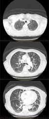
An unusual radiological presentation of pulmonary Langerhans' cell histiocytosis
- PMID: 22761212
- PMCID: PMC4543071
- DOI: 10.1136/bcr-03-2012-5980
An unusual radiological presentation of pulmonary Langerhans' cell histiocytosis
Abstract
Langerhans' cell histiocytosis is a proliferative disease of the dendritic cell lineage. It usually has a characteristic radiological presentation of apical nodules and small cysts occurring predominantly in young adult smokers. Here we report a case of multisystem Langerhans cell histiocytosis in a 75-year-old woman with unusual chest radiology.
Conflict of interest statement
Figures


Similar articles
-
[A case of pulmonary Langerhans' cell histiocytosis].Nihon Kokyuki Gakkai Zasshi. 2003 Sep;41(9):685-90. Nihon Kokyuki Gakkai Zasshi. 2003. PMID: 14531308 Japanese.
-
Pulmonary Langerhans' cell histiocytosis: radiologic resolution following smoking cessation.Chest. 1999 May;115(5):1452-5. doi: 10.1378/chest.115.5.1452. Chest. 1999. PMID: 10334170
-
A case of pulmonary langerhans' cell histiocytosis mimicking hematogenous pulmonary metastases.Korean J Intern Med. 2009 Dec;24(4):393-6. doi: 10.3904/kjim.2009.24.4.393. Epub 2009 Nov 27. Korean J Intern Med. 2009. PMID: 19949741 Free PMC article.
-
[Pneumothorax secondary to pulmonary histiocytosis X].Minerva Chir. 1999 Jul-Aug;54(7-8):531-6. Minerva Chir. 1999. PMID: 10528489 Review. Italian.
-
Pulmonary Langerhans' cell histiocytosis in adults.Adv Respir Med. 2017;85(5):277-289. doi: 10.5603/ARM.a2017.0046. Epub 2017 Oct 30. Adv Respir Med. 2017. PMID: 29083024 Review.
References
-
- Badalian-Very G, Vergilio J, Degar BA, et al. Recent advances in the understanding of Langerhans cell Histiocytosis. Br J Haematol 2012;156:163–72. - PubMed
-
- Medoff BD, Abbott GF, Louissant A. Case 16-2010: a 48 year old man with a cough and pain in the left shoulder. N Engl J Med 2010;362:2013–22. - PubMed
-
- Vassallo R, Ryu JH. Pulmonary Langerhans’ cell histiocytosis. Clin Chest Med 2004;25:561–71. - PubMed
-
- Vassallo R, Ryu JH, Schroeder DR, et al. Clinical outcomes of pulmonary Langerhans cell histiocytosis in adults. N Engl J Med 2002;346:484–90. - PubMed
-
- Kulwiec EL, Lynch DA, Aguaya SM, et al. imaging of pulmonary histiocytosis X. Radiographics 1992;12:515–26. - PubMed
Publication types
MeSH terms
Substances
LinkOut - more resources
Full Text Sources
Medical
