. 2012 Sep;214(3):421-35.
doi: 10.1530/JOE-12-0110. Epub 2012 Jul 3.

Duplicated zebrafish co-orthologs of parathyroid hormone-related peptide (PTHrP, Pthlh) play different roles in craniofacial skeletogenesis

Affiliations

Duplicated zebrafish co-orthologs of parathyroid hormone-related peptide (PTHrP, Pthlh) play different roles in craniofacial skeletogenesis

Yi-Lin Yan et al. J Endocrinol. 2012 Sep.

Abstract

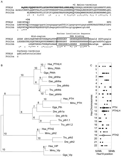
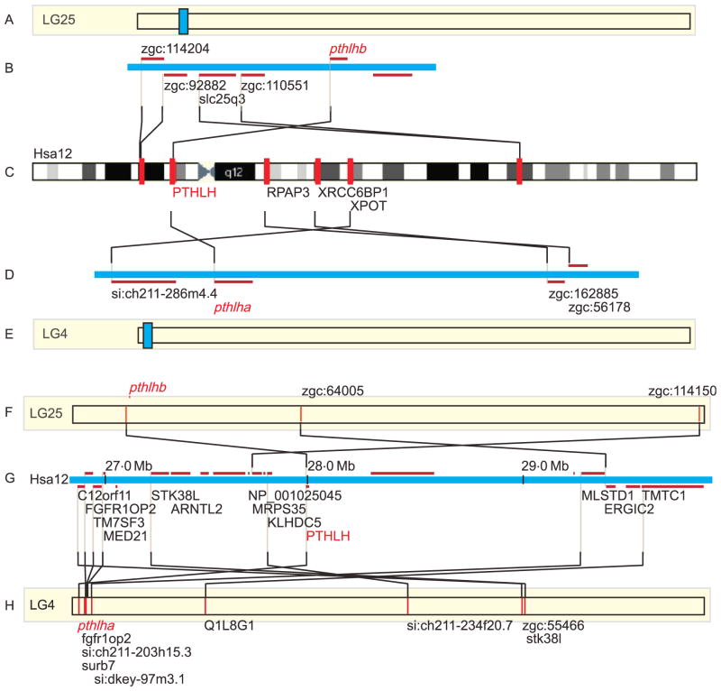
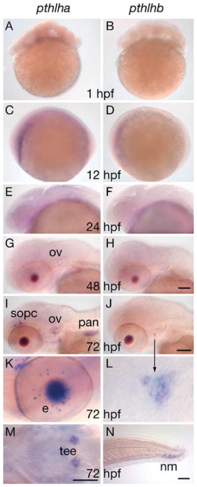
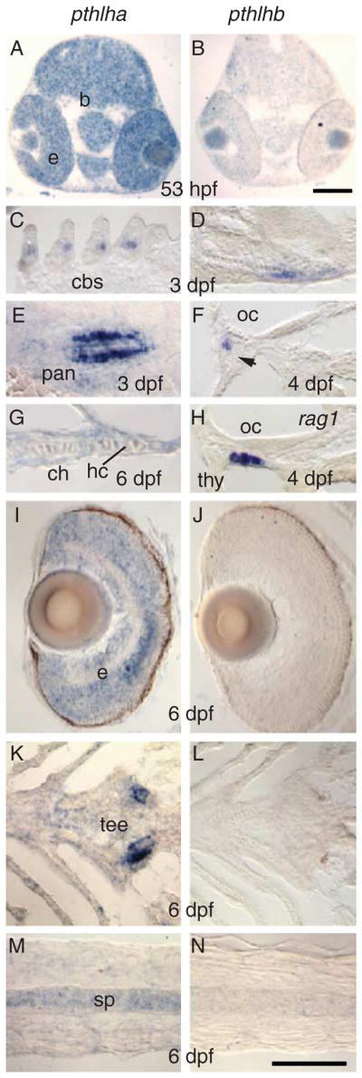
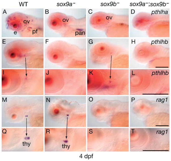
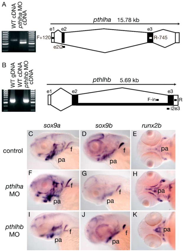
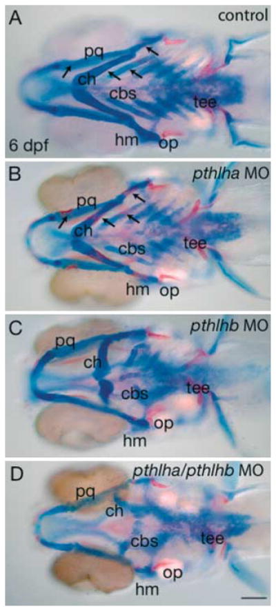
In mammals, parathyroid hormone-related peptide (PTHrP, alias PTH-like hormone (Pthlh)) acts as a paracrine hormone that regulates the patterning of cartilage, bone, teeth, pancreas, and thymus. Beyond mammals, however, little is known about the molecular genetic mechanisms by which Pthlh regulates early development. To evaluate conserved pathways of craniofacial skeletogenesis, we isolated two Pthlh co-orthologs from the zebrafish (Danio rerio) and investigated their structural, phylogenetic, and syntenic relationships, expression, and function. Results showed that pthlh duplicates originated in the teleost genome duplication. Zebrafish pthlha and pthlhb were maternally expressed and showed overlapping and distinct zygotic expression patterns during skeletal development that mirrored mammalian expression domains. To explore the regulation of duplicated pthlh genes, we studied their expression patterns in mutants and found that both sox9a and sox9b are upstream of pthlha in arch and fin bud cartilages, but only sox9b is upstream of pthlha in the pancreas. Morpholino antisense knockdown showed that pthlha regulates both sox9a and sox9b in the pharyngeal arches but not in the brain or otic vesicles and that pthlhb does not regulate either sox9 gene, which is likely related to its highly degraded nuclear localization signal. Knockdown of pthlha but not pthlhb caused runx2b overexpression in craniofacial cartilages and premature bone mineralization. We conclude that in normal cartilage development, sox9 upregulates pthlh, which downregulates runx2, and that the duplicated nature of all three of these genes in zebrafish creates a network of regulation by different co-orthologs in different tissues.


Conflict of interest statement

Declaration of interest

The authors declare that there is no conflict of interest that could be perceived as prejudicing the impartiality of the research reported.

Figures

Figure 1
Figure 1
Phylogenetic and conserved synteny analysis. (A) Human (M57293 and M32740) and zebrafish Pthlha (DQ022615) and Pthlhb (DQ022616) amino acid sequences aligned (upper case) as described (Bhattacharya et al. 2011). The first residue of human PTHLH is designated as +1 (underlined). The signal sequence and ‘pro’ peptides (residues to the left of +1) are bold. The Pthlh sequence alignments revealed a 41 amino acid gap of low sequence homology plus conserved regions including the N-terminus (1–34; 34th residue is underlined), nuclear localization sequence (boxed) containing the RNA-binding domain, and mid-region (uppercase italics). Because the zebrafish genome contains two Pthlh orthologs that differ in their conservation in these regions, they are useful natural variants to evaluate the roles different domains play during chondrogenic and osteogenic development. *Identical residues: conservative substitutions (Flanagan et al. 2000). (B) Phylogenetic analysis by maximum likelihood, maximum parsimony, and neighbor joining methods agreed in topology (maximum likelihood shown using JTT substitution model, four substitution rate categories, http://atgc.lirmm.fr/phyml/(Guindon et al. 2005). Bootstrap values are from ten replicates. Analyses suggest that PTH and Pthlh are sister groups and that PTH2 (alias TIP39) is the sister group to this super-clade. PTH, Pthlh, and PTH2 thus form a superfamily, whereas secretin falls as a sister group to this larger superfamily. (C) Dotplots mapping genes on Hsa19 from 52 to 58 Mb displayed along the horizontal axis to their orthologs on the other human chromosomes displayed as a dot on the proper chromosome along the vertical axis directly above each Hsa19 gene. Circles indicate the locations of PTH2 on Hsa19 and its paralogs PTH and PTHLH on human chromosomes Hsa11 and Hsa12 respectively. The PTH2 chromosome segment has paralogs mainly on Hsa11 and Hsa12. Tetrapods: chicken (Gallus gallus), Gga_Pth_M36522; human, Hsa_PTH_NM_000315; mouse, Mmu_Pth_NM_020623, Gga_Pthlh_X52131, Hsa_PTHLH_J03580, Mmu_Pthlh_M60056, Hsa_PTH2_NP_848544, Mmu_PTH2_NP_444486, Gga_Vip_AAA87896, Mmu_ Vip_BAB31301, VIP_Hsa_NP_003372; Fish: zebrafish (Danio rerio), Dre_PTH1a_NM_212950, Dre_PTH1b_NM_212949, fugu Tru_pth1.1 (Danks et al. 2003), Tru_pth1.2_AAQ73561; Tru_pthlhb_AJ249391; stickleback (Gasterosteus aculeatus), Gac_pthlhb_ENSGACG00000004317, Gac_pthlha_ENSGACT00000000765, pthlha_Dre_NP_001019798, pthlhb_Dre_NP_001036789, Tru_pth2_T004305 Scaffold_4305, Dre_pth2_NP_991140.
Figure 2
Figure 2
Conserved syntenies for zebrafish pthlha and pthlhb genes. (A, E, F and H) Represent entire zebrafish chromosomes (Zv7). (B and D) Show all annotated genes (red horizontal lines) in the regions of the zebrafish chromosomes indicated in blue in A and E. (C) Shows an ideogram of human chromosome 12, with the locations of human orthologs of zebrafish genes shown as red bars. Lines link orthologs between the two species. (G) Shows the region of human chromosome 12p11.2 with all annotated genes indicated in red horizontal lines. Annotated sequences for which no zebrafish ortholog is called are in gray. Black lines link human genes to zebrafish orthologs. The ortholog of ARNTL2 is on LG18.
Figure 3
Figure 3
Tissue-specific expression of zebrafish pthlh co-orthologs. Expression of pthlha and pthlhb assessed by whole-mount in situ hybridization (Pthlha panels A, C, E, G, I, K and M; Pthlhb panels B, D, F, H, J, L and N) assessed at 1, 12, 24, 48, and 72 hpf. Panels A and B show very low level of expression at the embryo–yolk interface for both pthlh mRNAs. The pthlha gene was expressed in the otic vesicle (ov, panels G and I); super optic cartilages (sopc), pancreas (pan, panel I), retina (e, panel K), and teeth (tee, panel M). The pthlhb gene was expressed in the pharyngeal arch region (panels J and L, arrow) and urophysis (neuromasts) of the tail (nm, panel N). Panels A, B, C, D, E, F, G, H, I and J, lateral view, anterior toward the left. All scale bars, 100 μm.
Figure 4
Figure 4
Histological analysis of tissue-specific expression of zebrafish pthlh co-orthologs. Expression of pthlha and pthlhb transcripts in sections of zebrafish embryos (pthlha panels A, C, E, G, I, K and M; pthlhb panels B, D, F, H, J, L and N) at 53 hpf, 3, 4, and 6 dpf. Expression of pthlha was robust in the brain (b) and retina (e, A), ceratobranchial arch cartilages (cbs, C), pancreas endocrine cells (pan, E), hypertrophic chondrocytes (hc) of the ceratohyal cartilage (ch, G), eye (e, A, I), teeth (tee, K), and spinalcord (sp, M). Expression of pthlhb was slight in the eye (retina, B), chondrocytes at the posterior border of the hyosymplectic and opercular (Fig. 4D and F respectively), and the thymus region (thm, H; rag1-labeled cRNA probe, control); oc, opercular chondrocytes (F and H). All scale bars, 100 μm.
Figure 5
Figure 5
pthlh expression in sox9 mutants. Expression of pthlha, pthlhb, and rag1 in WT, sox9a and sox9b single mutants, and sox9a/sox9b double mutants in 4 dpf embryos. pthlha expression (A) was diminished in the pharyngeal arches, superoptic cartilages, and pectoral fin buds of sox9a (B) and sox9b mutants (C). In double mutants, most pthlha expression was eliminated (D). We conclude that sox9a regulates pthlha expression in many craniofacial cartilages and in the pectoral fin (sox9apthlha in chondrocytes) and that pthlha is downstream of sox9a in some tissues and downstream of sox9b in other tissues. pthlhb (and not pthlha) showed expression in the a distinctive domain about 20 cells in the pharyngeal arch region (E, I and G, K). pthlhb expression in that pharyngeal arch region is regulated by sox9a (F, J) while rag1 expression in thymus is regulated by sox9b (O, S). The arrows show the hybridized region in a more amplified image below. Thus for example, the triangular region on panel E is shown in panel I at a greater magnification. Similarly the rag1 expression in panel M is shown in panel Q at a greater magnification. e, retina; ov, otic vesicle; pan, pancreas; pf, pectoral fin; and thm, thymus. Anterior towards the left. Scale bars, 100 μm.
Figure 6
Figure 6
Morpholino knockdown of pthlha and pthlhb. (A) pthlha; left, pthlha amplicon size difference between cDNAs isolated from WT and MO-treated embryos, right, schematic indicating exons 1–3 (boxes), introns (lines), primers (forward, F+120 and reverse, R−745), and splice-blocking MO (e2i2). Compared with WT, the pthlha morphant showed a significantly decreased amplicon size due to the elimination of exon-2 from the pre-mRNA. (B) pthlhb; left, pthlhb amplicon size difference between WT and MO-treated embryos. Right, schematic indicating the location of exons 1–3, primers (forward, F-in and reverse, R) and splice-blocking MO (i2e3). Compared with WT gDNA, the pthlhb morphant showed a similar amplicon size indicating the lack of intron excision in the mRNA whereas WT cDNA yielded no amplicon. (C, D, E, F, G, H, I, J and K) Regulation of sox9 expression in pthlha knockdown embryos (C, F and I) and pthlhb knockdown embryos (D, G and J) at 3 dpf queried by in situ hybridization for sox9a, sox9b, and runx2b as indicated in the figure. C, D, F, G, I and J, lateral views. E, H and K, ventral views with anterior to the left. f, pectoral fin; pa, pharyngeal arches.
Figure 7
Figure 7
Regulation of pharyngeal arch development by Pthlh co-orthologs at 6 dpf. 6 Dpf fish stained with alcian blue and alizarin red. pthlha MO-treated embryos (B) showed significantly more premature alizarin red staining (bone formation, arrows) compared with noninjected controls (A). pthlhb MO-treated embryos (C) showed deformed pharyngeal skeletons compared with noninjected controls (A) and pthlha knockdown (B). Double knockdown of pthlha and pthlhb (D) showed severe abnormality in the pharyngeal skeleton. cbs, ceratobranchials; ch, ceratohyal; hm, hyomandibular; op, opercular; pq, palatoquadrate; tee, teeth. Scale bar is 100 μm.

Similar articles

Cited by

References

    1. Abbink W, Flik G. Minireview: parathyroid hormone-related protein in teleost fish. General and Comparative Endocrinology. 2007;152:243–251. doi: 10.1016/j.ygcen.2006.11.010. - DOI - PubMed
    1. Akiyama H, Kim J-E, Nakashima K, Balmes G, Iwai N, Deng J, Zhang Z, Martin J, Behringer R, Nakamura T, et al. Osteo-chondroprogenitor cells are derived from Sox9 expressing precursors. PNAS. 2005;102:14665–14670. doi: 10.1073/pnas.0504750102. - DOI - PMC - PubMed
    1. Albertson R, Payne-Fereira T, Postlethwait J, Yelick P. Zebrafish acvr2a and acvr2b exhibit distinct roles in craniofacial development. Developmental Dynamics. 2005;233:1405–1418. doi: 10.1002/dvdy.20480. - DOI - PubMed
    1. Alt B, Reibe S, Feitosa N, Elsalini O, Wendl T, Rohr K. Analysis of origin and growth of the thyroid gland in zebrafish. Developmental Dynamics. 2006;235:1872–1883. doi: 10.1002/dvdy.20831. - DOI - PubMed
    1. Amores A, Force A, Yan Y-L, Joly L, Amemiya C, Fritz A, Ho R, Langeland J, Prince V, Wang Y-K, et al. Zebrafish hox clusters and vertebrate genome evolution. Science. 1998;282:1711–1714. doi: 10.1126/science.282.5394.1711. - DOI - PubMed

Publication types

MeSH terms