. 2012 Oct;343(1):97-105.
doi: 10.1124/jpet.112.196626. Epub 2012 Jul 3.

Aldehyde dehydrogenase type 2 activation by adenosine and histamine inhibits ischemic norepinephrine release in cardiac sympathetic neurons: mediation by protein kinase Cε

Affiliations

Aldehyde dehydrogenase type 2 activation by adenosine and histamine inhibits ischemic norepinephrine release in cardiac sympathetic neurons: mediation by protein kinase Cε

Pablo A Robador et al. J Pharmacol Exp Ther. 2012 Oct.

Abstract

During myocardial ischemia/reperfusion, lipid peroxidation leads to the formation of toxic aldehydes that contribute to ischemic dysfunction. Mitochondrial aldehyde dehydrogenase type 2 (ALDH2) alleviates ischemic heart damage and reperfusion arrhythmias via aldehyde detoxification. Because excessive norepinephrine release in the heart is a pivotal arrhythmogenic mechanism, we hypothesized that neuronal ALDH2 activation might diminish ischemic norepinephrine release. Incubation of cardiac sympathetic nerve endings with acetaldehyde, at concentrations achieved in myocardial ischemia, caused a concentration-dependent increase in norepinephrine release. A major increase in norepinephrine release also occurred when sympathetic nerve endings were incubated in hypoxic conditions. ALDH2 activation substantially reduced acetaldehyde- and hypoxia-induced norepinephrine release, an action prevented by inhibition of ALDH2 or protein kinase Cε (PKCε). Selective activation of G(i/o)-coupled adenosine A(1), A(3), or histamine H(3) receptors markedly inhibited both acetaldehyde- and hypoxia-induced norepinephrine release. These effects were also abolished by PKCε and/or ALDH2 inhibition. Moreover, A(1)-, A(3)-, or H(3)-receptor activation increased ALDH2 activity in a sympathetic neuron model (differentiated PC12 cells stably transfected with H(3) receptors). This action was prevented by the inhibition of PKCε and ALDH2. Our findings suggest the existence in sympathetic neurons of a protective pathway initiated by A(1)-, A(3)-, and H(3)-receptor activation by adenosine and histamine released in close proximity of these terminals. This pathway comprises the sequential activation of PKCε and ALDH2, culminating in aldehyde detoxification and inhibition of hypoxic norepinephrine release. Thus, pharmacological activation of PKCε and ALDH2 in cardiac sympathetic nerves may have significant protective effects by alleviating norepinephrine-induced life-threatening arrhythmias that characterize myocardial ischemia/reperfusion.


Figures
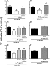
Fig. 1.
Fig. 1.
Release of NE from cardiac synaptosomes during 30-min hypoxia. Bars (means ± S.E.M.) represent the hypoxia-induced increase in NE release above the normoxic basal level of 273 ± 11.5 pmol/mg protein (n = 49). A, activation of ALDH2 with Alda-1 (20 μM; 12 min) reduces hypoxia-induced NE release. Desensitization of ALDH2 with GTN (2 μM; 30 min) prevents the effect of Alda-1 (n = 7–12). *, P < 0.05 versus hypoxia. #, P < 0.05 versus Alda-1 by unpaired t test. B, PKCε activation with ΨεRACK (PKCε activator; 500 nM; 12 min) reduces hypoxia-induced NE release. ALDH2 desensitization with GTN (2 μM; 30 min) prevents the effect of ΨεRACK (n = 7–8). *, P < 0.05 versus hypoxia. #, P < 0.05 versus ΨεRACK by unpaired t test. C, activation of adenosine A1 receptor with 2′-MeCCPA (A1R agonist; 10 nM; 12 min) diminishes hypoxia-induced NE release. ALDH2 desensitization with GTN (2 μM; 30 min) and PKCε blockade with εV1–2 (PKCε inhibitor; 1 μM; 20 min) each prevents the effect of 2′-MeCCPA (n = 7–10). **, P < 0.005 versus hypoxia. #, P < 0.05 versus 2′-MeCCPA by unpaired t test. D, blockade of adenosine A1-receptor with DPCPX (A1R antagonist; 300 nM; 12 min) enhances hypoxia-induced NE release (n = 15–17). *, P < 0.05 versus hypoxia by unpaired t test. E, selective activation of adenosine A3 receptor with IB-MECA (A3R agonist; 50 nM; 12 min) reduces hypoxia-induced NE release. ALDH2 desensitization with GTN (2 μM; 30 min) and PKCε blockade with εV1–2 (1 μM; 20 min) each prevents the effect of IB-MECA (n = 7–11). **, P < 0.01 versus hypoxia. ##, P < 0.005 versus IB-MECA. ###, P < 0.001 versus IB-MECA by unpaired t test. F, blockade of adenosine A3 receptor with MRS1523 (A3R antagonist; 100 nM; 12 min) enhances hypoxia-induced NE (n = 7–9). *, P < 0.05 versus hypoxia by unpaired t test.
Fig. 2.
Fig. 2.
Incubation of isolated cardiac synaptosomes with acetaldehyde elicits a concentration-dependent increase in NE release, which is inhibited by the activation of either ALDH2 or PKCε. Points (means ± S.E.M.; n = 4–46 and 12–24 in A and B, respectively) represent percentage increases in NE release above a mean basal control level of 205 ± 7.4 pmol/mg protein (n = 92). A, ALDH2 activation with Alda-1 (20 μM; 10 min) attenuates the acetaldehyde-induced release of NE. ALDH2 desensitization with GTN (2 μM; 30 min) prevents the effect of Alda-1. B, PKCε activation with ΨεRACK (500 nM, 10 min) attenuates the release of NE. ALDH2 desensitization with GTN (2 μM; 30 min) prevents the effect of ΨεRACK. ***, P < 0.0001 from control and GTN + Alda-1 in A and from control and GTN + ΨεRACK in B, by unpaired t test.
Fig. 3.
Fig. 3.
Activation of adenosine A1 and A3 receptors inhibits the acetaldehyde-induced NE release from isolated cardiac synaptosomes: prevention by either PKCε inhibition or ALDH2 desensitization. Points (means ± S.E.M.; n = 8–42) represent percentage increases in NE release above a mean basal control level of 267 ± 6.6 pmol/mg protein (n = 124). A, selective activation of A1 receptors with 2′-MeCCPA (10 nM; 10 min) attenuates NE release by acetaldehyde, an action that is prevented by A1-receptor blockade with DPCPX (300 nM; 10 min). B and C, the A1 receptor-induced attenuation of NE release is prevented either by PKCε inhibition with εV1–2 (1 μM; 10 min) (B) or ALDH2 desensitization with GTN (2 μM; 30 min) (C). D, selective activation of A3 receptors with IB-MECA (50 nM; 10 min) attenuates NE release by acetaldehyde, an action that is prevented by A3-receptor blockade with MRS1523 (100 nM; 10 min). E and F, the A3 receptor-induced attenuation of NE release is prevented either by PKCε inhibition with εV1–2 (1 μM; 10 min) (E) or ALDH2 desensitization with GTN (2 μM; 30 min) (F). **, P < 0.01 and ***, P < 0.001 from control and A1- and A3-receptor agonists in combination with respective antagonists or in combination with εV1–2 or GTN, by unpaired t test.
Fig. 4.
Fig. 4.
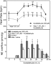
ALDH2 activation inhibits the positive chronotropic effect of acetaldehyde and associated increase in NE overflow in guinea pig hearts ex vivo. Top, time course of the increase in spontaneous heart rate during perfusion with acetaldehyde (500 μM). Pretreatment with the ALDH2 activator Alda-1 (20 μM; 10 min) inhibits the acetaldehyde-induced tachycardia and attenuates the associated increase in NE overflow. Points are heart rates recorded at the corresponding times on the abscissa (means ± S.E.M.; n = 5 and 6 for control and Alda-1, respectively). †, P < 0.05 and ††, P <0.01 from control by one-way ANOVA + Bonferroni's test. **, P < 0.01 and ***, P < 0.001 from acetaldehyde, by two-way ANOVA + Bonferroni's test. Bottom, time course of the increase in NE overflow (measured in 5-min intervals) in the same hearts as in Top. Bars are means (means ± S.E.M.; n = 5 and 6 for control and Alda-1, respectively). †, P < 0.05 and ††, P < 0.01 from control by one-way ANOVA + Bonferroni's test. **, P < 0.01 from acetaldehyde, by two-way ANOVA + Bonferroni's test.
Fig. 5.
Fig. 5.
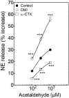
Incubation of isolated cardiac synaptosomes with acetaldehyde elicits a concentration-dependent increase in NE release that is potentiated by the NE transporter inhibitor DMI (300 nM; 10 min). A selective N-type Ca2+ channel blocker, ω-CTX (100 nM; 10 min), markedly reduces the acetaldehyde-induced NE release. Points (means ± S.E.M.; n = 8–32) represent percentage increases in NE release above a mean basal control level of 246 ± 6.80 pmol/mg protein (n = 32). ***, P < 0.001 from acetaldehyde control by unpaired t test.
Fig. 6.
Fig. 6.
Release of NE from cardiac synaptosomes during 30-min hypoxia. Bars (means ± S.E.M.) represent the hypoxia-induced increase in NE release above the normoxic basal level of 291 ± 24 pmol/mg protein (n = 14). Treatment with the selective histamine H3-receptor agonist methimepip (1 nM; 12 min) reduces hypoxia-induced NE release, and this effect is blocked by a previous addition of the selective histamine H3-receptor antagonist JNJ5207852 (30 nM; 12 min). PKCε blockade with εV1–2 (1 μM; 20 min) and ALDH2 desensitization with GTN (2 μM; 30 min) each prevents the effect of methimepip (n = 5–16). ***, P < 0.001 versus hypoxia. ##, P < 0.005 versus methimepip. ###, P < 0.0005 versus methimepip by unpaired t test.
Fig. 7.
Fig. 7.
Activation of histamine H3 receptors inhibits the acetaldehyde-induced NE release from isolated cardiac synaptosomes: prevention by either PKCε inhibition or ALDH2 desensitization. Points (means ± S.E.M.; n = 12–28) represent percentage increases in NE release above a mean basal control level of 268 ± 9.2 pmol/mg protein (n = 96). A, selective activation of H3 receptors with methimepip (1 nM; 10 min) attenuates NE release by acetaldehyde, an action that is prevented by H3-receptor blockade with JNJ5207852 (30 nM; 10 min). B and C, the H3-receptor-induced attenuation of NE release is prevented either by PKCε inhibition with εV1–2 (1 μM; 10 min) (B) or ALDH2 desensitization with GTN (2 μM; 30 min) (C). ***, P < 0.0001 from control and H3-receptor agonists with respective antagonists or εV1–2 and GTN, by unpaired t test.
Fig. 8.
Fig. 8.
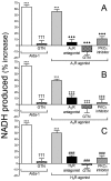
Activation of adenosine A1 receptors, A3 receptors, or histamine H3 receptors increases ALDH2 activity in PC12-H3 cells. Incubation of PC12-H3 cells with the selective ALDH2 activator Alda-1 (100 μM; 20 min) increases ALDH2 activity (measured by the rate of NADH production at 340 nm). A, incubation of PC12-H3 cells with the A1-receptor agonist 2′-MeCCPA (10 nM; 20 min) increases ALDH2 activity. Selective desensitization of ALDH2 with GTN (2 μM; 30 min) prevents the effects of Alda-1 and A1-receptor agonist. Pretreatment of PC12-H3 cells with the selective A1-receptor antagonist DPCPX (300 nM; 30 min) or the selective PKCε inhibitor εV1–2 (1 μM; 30 min) prevents the effects of A1-receptor activation. B, incubation of PC12-H3 cells with the A3-receptor agonist IB-MECA (50 nM; 20 min) increases ALDH2 activity. Selective desensitization of ALDH2 with GTN (2 μM; 30 min) prevents the effects of Alda-1 and IB-MECA. Pretreatment of PC12-H3 cells with the selective A3-receptor antagonist MRS1523 (100 nM; 30 min) or the selective PKCε inhibitor εV1–2 (1 μM; 30 min) prevents the effects of A3-receptor activation. C, incubation of PC12-H3 cells with the H3-receptor agonist methimepip (1 nM; 20 min) increases ALDH2 activity. Selective desensitization of ALDH2 with GTN (2 μM; 30 min) prevents the effects of Alda-1 and methimepip. Pretreatment of PC12-H3 cells with the selective H3-receptor antagonist JNJ5207852 (30 nM; 30 min) or the selective PKCε inhibitor εV1–2 (1 μM; 30 min) prevents the effects of H3-receptor activation. Bars are mean percentage increases from control (± S.E.M.; n = 4–16). Basal NADH production was 7.47 ± 0.13 μmol/min/mg protein. ***, P < 0.0001 from control. †††, P < 0.0001 from Alda-1. ♦♦♦, P < 0.0001 from A1R agonist. ‡‡‡, P < 0.001 from A3R agonist. ###, P < 0.0001 from methimepip by unpaired t test.
Fig. 9.
Fig. 9.
Proposed pathway for the inhibition of NE release from cardiac sympathetic neurons upon activation of mitochondrial ALDH2. This involves the pivotal phosphorylation/activation of mitochondrial ALDH2 by PKCε, which is translocated/activated when Gi/o-coupled receptors, such as A1, A3, and H3 receptors, are activated by adenosine and histamine released in ischemic conditions in close proximity to sympathetic nerve endings.

Similar articles

Cited by

References

    1. Aloyo VJ, McIlvain HB, Bhavsar VH, Roberts J. (1991) Characterization of norepinephrine accumulation by a crude synaptosomal-mitochondrial fraction isolated from rat heart. Life Sci 48:1317–1324 - PubMed
    1. Baraldi PG, Tabrizi MA, Fruttarolo F, Romagnoli R, Preti D. (2008) Recent improvements in the development of A2B adenosine receptor agonists. Purinergic Signal 4:287–303 - PMC - PubMed
    1. Barbier AJ, Berridge C, Dugovic C, Laposky AD, Wilson SJ, Boggs J, Aluisio L, Lord B, Mazur C, Pudiak CM, et al. (2004) Acute wake-promoting actions of JNJ-5207852, a novel, diamine-based H3 antagonist. Br J Pharmacol 143:649–661 - PMC - PubMed
    1. Carini R, De Cesaris MG, Spendore R, Albano E. (2000) Ethanol potentiates hypoxic liver injury: role of hepatocyte Na+ overload. Biochim Biophys Acta 1502:508–514 - PubMed
    1. Chen CH, Budas GR, Churchill EN, Disatnik MH, Hurley TD, Mochly-Rosen D. (2008) Activation of aldehyde dehydrogenase-2 reduces ischemic damage to the heart. Science 321:1493–1495 - PMC - PubMed

Publication types

MeSH terms

LinkOut - more resources