. 2012 Jul 31;109(31):12770-5.
doi: 10.1073/pnas.1202944109. Epub 2012 Jul 3.

Modeling human cortical development in vitro using induced pluripotent stem cells

Affiliations

Modeling human cortical development in vitro using induced pluripotent stem cells

Jessica Mariani et al. Proc Natl Acad Sci U S A. .

Abstract

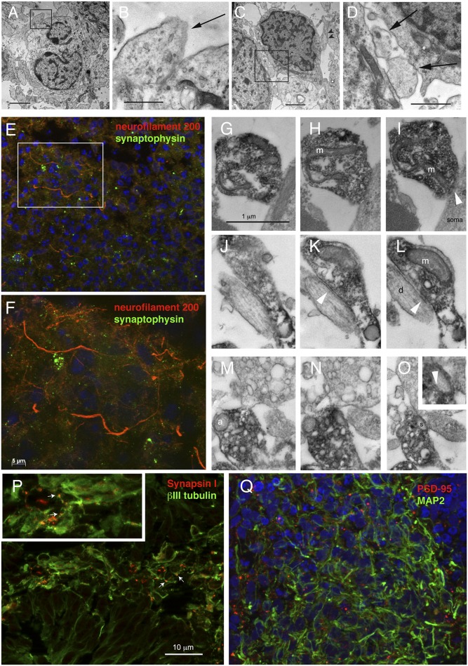
Human induced pluripotent stem cells (hiPSCs) are emerging as a tool for understanding human brain development at cellular, molecular, and genomic levels. Here we show that hiPSCs grown in suspension in the presence of rostral neuralizing factors can generate 3D structures containing polarized radial glia, intermediate progenitors, and a spectrum of layer-specific cortical neurons reminiscent of their organization in vivo. The hiPSC-derived multilayered structures express a gene expression profile typical of the embryonic telencephalon but not that of other CNS regions. Their transcriptome is highly enriched in transcription factors controlling the specification, growth, and patterning of the dorsal telencephalon and displays highest correlation with that of the early human cerebral cortical wall at 8-10 wk after conception. Thus, hiPSC are capable of enacting a transcriptional program specifying human telencephalic (pallial) development. This model will allow the study of human brain development as well as disorders of the human cerebral cortex.


Conflict of interest statement

The authors declare no conflict of interest.

Figures

Fig. 1.
Fig. 1.
Radial glia and excitatory progenitors differentiate from hiPSCs at day 50 in vitro. Day-50 forebrain-like structures contain radial glial cells immunoreactive for NESTIN, PAX6, and BLBP (A, B, and E), and βIIITUBULIN+ and TBR1+ neurons (B, D, and G). The radial glia express SOX1 and SOX2 (H–L), are mitotically active, as shown by Ki67 expression (M–O, red), and are polarized, displaying N-CADHERIN+ apical end-feet (C and F) as well as apical mitoses (M–O, arrowheads). A layer of TBR2+ intermediate progenitors (C and F) surrounds the radial glial layer and displays pH3+ basal mitoses (M–O, arrows). The neurons express MAP2 but rarely ZIC1 (P and Q) and do not express ISLET1 and OLIG2 (R). (Scale bars, 200 μm in A–D and M; 5 μm in E–G and I; 10 μm in H, J–L, N, and O.)
Fig. 2.
Fig. 2.
Evidence for synapses in hiPSC-derived multilayered structures at 50 and 70 d in vitro. (A–D) Day-70 multilayered structures analyzed by EM show examples of direct apposition between vesicles-containing putative axon terminals (black arrows) and neuronal cell bodies. (E and F) Synaptophysin/neurofilament 200 double immunostaining at day 70 showing numerous synaptophysin-labeled boutons decorating the neurites. (G–O) Immunoelectron microscopy showing synaptophysin-immunoprecipitate within putative axon terminals contacting cell bodies (soma) (G–I), dendrites (d) (J–L), and spines (s) (M–O). White arrowheads, synaptic thickening; m, mitochondia; a, autophagosome. (P and Q) Day-50 multilayered structures stained for SYN I and βIIITUBULIN (P) or PSD-95 and MAP2 (Q). White arrows in P point to SYN I-positive boutons. (Scale bars, 2 μm in A and C; 0.5 μm in B and D; 10 μm in E, P, and Q; 5 μm in F; 1 μm in G–O.)
Fig. 3.
Fig. 3.
Regional and temporal specification of hiPSC-derived neuronal cells. (A) Fold differences in neuronal gene expression compared with undifferentiated hiPSCs for dorsally and ventrally enriched telencephalic genes. The gene lists were manually curated according to published data (24, 25, 35). Orange, genes that are statistically different at P < 0.05 and fold change ±2.0; gray, genes that do not fulfill the minimum P value criterion. (B) Number of developing human brain samples in Kang et al. (4) correlating with the day-50 multilayered structures within 95% confidence interval of the maximal correlation. Human brain samples are displayed according to developmental stages (Upper) or brain regions (Lower). PCW, postconceptional week; M, months; Y, years; FC, frontal, PC, parietal, TC, temporal, and OC, occipital cerebral cortical wall; HIP, hippocampal anlage; AMY, amygdala; DIE, diencephalon; URL, upper rhombic lip.
Fig. 4.
Fig. 4.
Specification of cortical layer fates in hiPSC-derived neurons. (A) Scheme of TF directing cortical layer fates. (B–R) Immunostaining for TFs in sections from 3D structures at day 50 in vitro. (B) Double immunostaining for MAP2 and CTIP2, showing CTIP2 nuclear localization in MAP2+ cells; (C–F) TBR1 and CTIP2; (G–I) TBR1 and SATB2; (J–M) TLE4 and CTIP2; (N–P) TLE4 and SATB2; and (Q and R) TBR1 and BRN2, showing differences in relative colocalization of these TFs in differentiating neurons. White arrowheads, cells stained for a single TF; white arrows, cells that colocalize two factors. Broken line in Q delimits the radial glial layer where BRN2 is also expressed but was excluded from quantification. (S) Stereological quantification of TF expression in 3D structures cultured with different amounts of FGF2 for the first 18 d. DAPI is in blue. (Scale bars, 200 μm in B and G; 100 μm in C–F and J; 25 μm in H and I; 20 μm in K–P.)

References

    1. Rakic P. Evolution of the neocortex: A perspective from developmental biology. Nat Rev Neurosci. 2009;10:724–735. - PMC - PubMed
    1. Geschwind DH. Neurodevelopmental disorders: Hope for a new beginning. Curr Opin Neurol. 2011;24:95–97. - PMC - PubMed
    1. Johnson MB, et al. Functional and evolutionary insights into human brain development through global transcriptome analysis. Neuron. 2009;62:494–509. - PMC - PubMed
    1. Kang HJ, et al. Spatio-temporal transcriptome of the human brain. Nature. 2011;478:483–489. - PMC - PubMed
    1. Brennand KJ, et al. Modelling schizophrenia using human induced pluripotent stem cells. Nature. 2011;473:221–225. - PMC - PubMed

Publication types

MeSH terms

Substances