. 2012 Sep;86(18):9888-98.
doi: 10.1128/JVI.00956-12. Epub 2012 Jul 3.

Interferon response factors 3 and 7 protect against Chikungunya virus hemorrhagic fever and shock

Affiliations

Interferon response factors 3 and 7 protect against Chikungunya virus hemorrhagic fever and shock

Penny A Rudd et al. J Virol. 2012 Sep.

Abstract

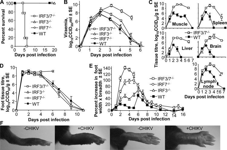
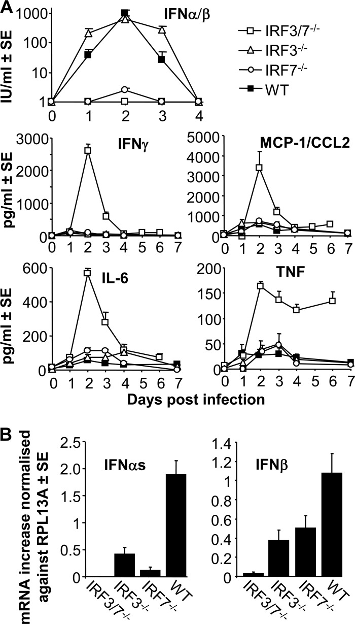
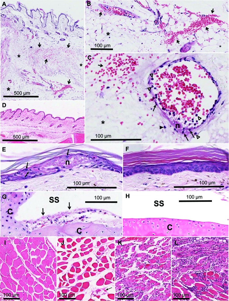
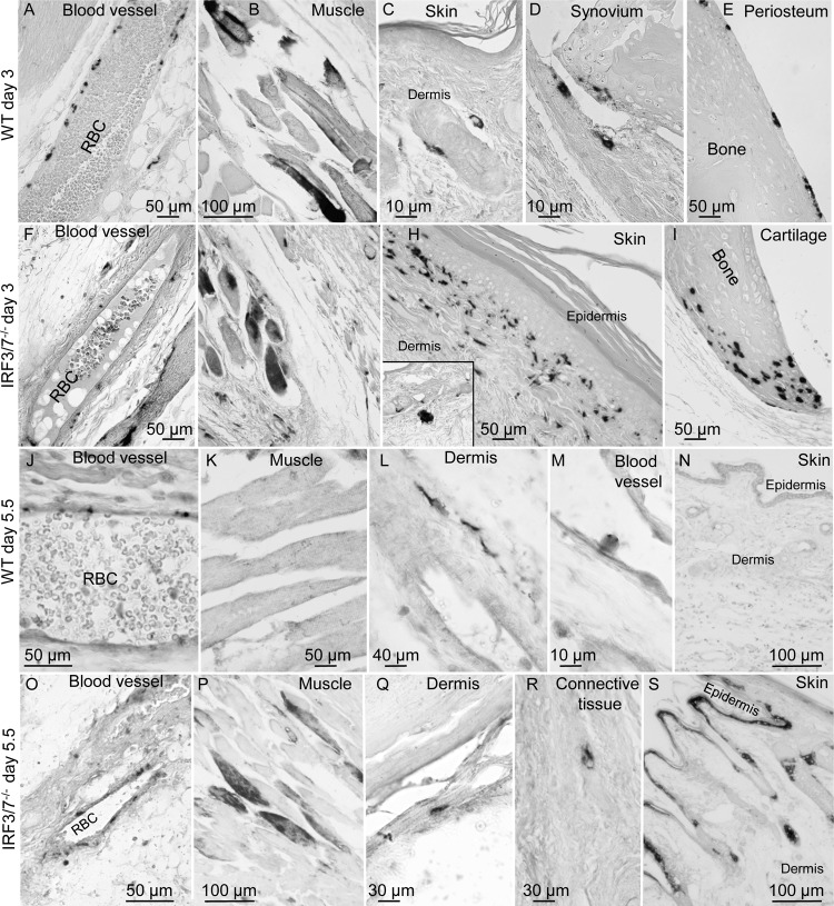
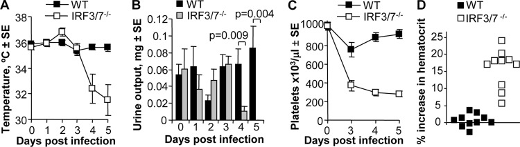
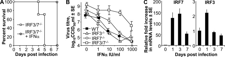
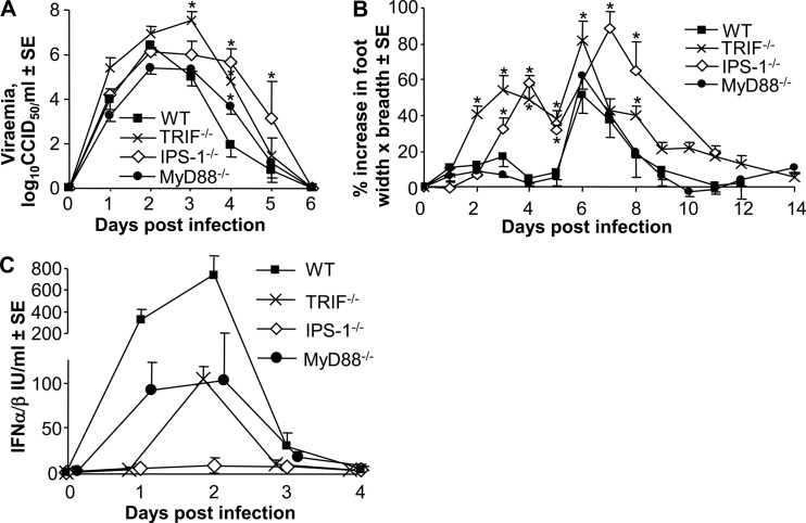
Chikungunya virus (CHIKV) infections can produce severe disease and mortality. Here we show that CHIKV infection of adult mice deficient in interferon response factors 3 and 7 (IRF3/7(-/-)) is lethal. Mortality was associated with undetectable levels of alpha/beta interferon (IFN-α/β) in serum, ∼50- and ∼10-fold increases in levels of IFN-γ and tumor necrosis factor (TNF), respectively, increased virus replication, edema, vasculitis, hemorrhage, fever followed by hypothermia, oliguria, thrombocytopenia, and raised hematocrits. These features are consistent with hemorrhagic shock and were also evident in infected IFN-α/β receptor-deficient mice. In situ hybridization suggested CHIKV infection of endothelium, fibroblasts, skeletal muscle, mononuclear cells, chondrocytes, and keratinocytes in IRF3/7(-/-) mice; all but the latter two stained positive in wild-type mice. Vaccination protected IRF3/7(-/-) mice, suggesting that defective antibody responses were not responsible for mortality. IPS-1- and TRIF-dependent pathways were primarily responsible for IFN-α/β induction, with IRF7 being upregulated >100-fold in infected wild-type mice. These studies suggest that inadequate IFN-α/β responses following virus infection can be sufficient to induce hemorrhagic fever and shock, a finding with implications for understanding severe CHIKV disease and dengue hemorrhagic fever/dengue shock syndrome.


Figures

Fig 1
Fig 1
CHIKV infection in WT, IRF3−/−, IRF7−/−, and IRF3/7−/− mice. Mice (6 to 8 weeks old) were infected with CHIKV. (A) Mice were monitored daily, and a death event was recorded when an animal died or when animal welfare considerations required euthanasia (n = 12 to 20 per group; data are derived from three or four independent experiments). (B) Peripheral blood viremia (n = 7 to 12 per group; data are derived from three or four independent experiments). Differences between WT and IRF3/7−/− mice on days 3, 4, and 5 were significant (P < 0.001, P = 0.004, and P = 0.014, respectively; Mann-Whitney U test). (C) Tissue virus titers (n = 3 or 4 per group). Differences between WT and IRF3/7−/− mice were significant on days 3 and 4 (except for spleen on day 4) (P < 0.05; Mann-Whitney U test). (D) Virus titers in feet (n = 3 to 5 per group). Differences between WT and IRF3/7−/− mice reached significance on day 6 (P = 0.01, Mann-Whiney U test). (E) Percentage increase in foot swelling compared with day 0 (n = 30 feet per group, except for IRF3/7−/− day 6, where n = 6 feet). (Data are derived from three or four independent experiments.) (F) Photographs of feet from IRF3/7−/− mice before CHIKV infection (-CHIKV) and on day 4 postinfection (+CHIKV).
Fig 2
Fig 2
Cytokine and chemokine levels in the peripheral blood of CHIKV-infected mice. (A) WT, IRF3−/−, IRF7−/−, and IRF3/7−/− mice were infected with CHIKV, and peripheral blood samples were taken at the indicated times and assayed for the levels of the indicated cytokine or chemokine. The detection limit for the IFN-α/β bioassay was ∼1 IU/ml. The levels in IRF3/7−/− mice were significantly different from those in WT mice on days 2 and 3 for all cytokines and chemokines, on day 1 for IFN-α/β, and on day 4 for TNF (P < 0.034, Mann-Whitney U test). Day 2 IFN-α/β levels were also significantly higher in IRF7−/− mice than in IRF3/7−/− mice (P < 0.008, Mann-Whitney U test) (n = 4 to 6 per group). (B) Quantitative real-time RT-PCR for IFN-αs and IFN-β mRNAs in feet on day 2 postinfection. Data were normalized to RPL13A (housekeeping gene) (15), and values for uninfected feet were subtracted (n = 6 to 8 per group). All differences were significant (P < 0.03) except that for IFN-β in IRF3−/− and IRF7−/− mice.
Fig 3
Fig 3
Histology of IRF3/7−/− mice after CHIKV infection. (A) Subcutaneous foot tissue 5 days after infection with CHIKV showing severe generalized edema (asterisks) and severe multifocal hemorrhage (arrows). (B) Enlargement of bottom of panel A showing edema (asterisk) and hemorrhage (arrows). (C) Vasculitis on day 5 characterized by vascular wall fibrinoid necrosis (n) and intramural degenerating leukocytes (arrows). Some neutrophils (white arrowheads) and mononuclear cells (black arrowheads) are present; an enlarged image is shown in File S5 in the supplemental material. (D) Subcutaneous foot tissue of an uninfected IRF3/7−/− mouse. (E) Focal skin necrosis (n) 5 days after infection of IRF3/7−/− mice with CHIKV. Keratinocytes undergoing ballooning degeneration, with pale cytoplasm and karyorrhectic nuclei (arrows), are evident. (F) Skin of uninfected IRF3/7−/− mice. (G) Exudative arthritis evident in the joints of an IRF3/7−/− mouse 5 days postinfection with CHIKV. Inflammatory cells and fibrin are present (arrows) in the synovial space (SS). Articular cartilage (C) is indicated. (H) Articular cartilage from an uninfected IRF3/7−/− mouse. Labels are as in panel G. (I) Skeletal muscle of an uninfected IRF3/7−/− mouse. (J) Skeletal muscle of an IRF3/7−/− mouse on day 5 postinfection. (K) Skeletal muscle of an IRF7−/− mouse on day 5 postinfection. (L) Skeletal muscle of an IRF3−/− mouse on day 5 postinfection.
Fig 4
Fig 4
Detection of CHIKV RNA by in situ hybridization. CHIKV RNA was detected by in situ hybridization in tissues from CHIKV-infected (i) WT mice 3 days postinfection (A to E), (ii) IRF3/7−/− mice 3 days postinfection (F to I), (iii) WT mice 5.5 days postinfection (J to N), and (iv) IRF3/7−/− mice 5.5 days postinfection (O to S). Staining is shown for cells lining blood vessel walls (A, F, J, and O) (RBC, red blood cells), skeletal muscle (B, G, K, and P), skin (C, H, N, and S), synovial membrane (D), periosteum (E), articular cartilage (I), dermis (L and Q), a cell within a blood vessel (M), and connective tissue (R). Enlarged images of selected cells with more detailed descriptions are shown in File S6 in the supplemental material.
Fig 5
Fig 5
Clinical observations. (A) Body temperatures measured by a thermocouple placed in the pit of the hind leg (n = 4 to 16 per group). IRF3/7−/− mice showed a significant fever on day 2 (P = 0.004 for day 0 versus day 2). Temperatures were also higher in IRF3/7−/− mice than WT mice on day 2 (P = 0.01). Temperatures were significantly lower in IRF3/7−/− mice than WT mice on days 4 and 5 (P < 0.001). (B) Scruff-induced urine output. Differences were significant on days 4 and 5 (n = 8 to 10 per group). (C) Platelet count. Platelet counts were significantly lower in IRF7−/− than WT mice on days 3, 4, and 5 postinfection (P = 0.001, P < 0.001, and P < 0.001, respectively; t test; n = 8 to 10 per group). (D) Percentage increase in hematocrit in WT and IRF3/7−/− mice on day 5 postinfection compared with control uninfected mice (P < 0.001; n = 10 or 11 per group). All statistics were obtained by the Mann-Whitney U test. The data were obtained from two to four independent experiments.
Fig 6
Fig 6
IFN-α treatment and IRF7 induction. (A) IRF3/7−/− mice (n = 3) were treated with 104 IU of IFN-α i.v. 1 day before infection with CHIKV, and survival was monitored over time (IRF3/7−/− + IFN-α) and compared with that infected IRF3/7−/− mice not receiving IFN-α (n = 24) (log rank test, P = 0.001). (B) MEFs from IRF3/7−/−, IRF3−/−, IRF7−/−, and WT mice were treated in vitro with the indicated concentration of IFN-α overnight prior to CHIKV infection (MOI = 0.1). After washing, the cells were cultured for 24 h, and the supernatants were then assayed for viral titers (n = 3). (C) Quantitative real-time RT-PCR of IRF3 and IRF7 mRNA expression in CHIKV-infected feet normalized to RPL13A (n = 6). Levels are expressed relative to those on day 0.
Fig 7
Fig 7
CHIKV infection of IPS-1−/−, MyD88−/− and TRIF−/− mice. Viremia, foot swelling, and serum IFN-α/β levels following CHIKV infection of WT, TRIF−/−, IPS-1−/−, and MyD88−/− mice (8 to 12 weeks old). (A) Viremia. Significant differences between the knockout and WT mice are indicated (*). TRIF−/− mice (n = 11) showed higher viremias on days 3 and 4 than WT mice (n = 21 to 25; P = 0.001 and 0.004, respectively). IPS-1−/− mice (n = 3) showed higher viremias on days 4 and 5 than WT mice (P = 0.029 and 0.048, respectively). MyD88−/− (n = 8) mice showed a higher viremia on day 4 (P = 0.013). (B) Foot swelling. TRIF−/− feet (n = 10 to 32) were significantly larger on days 2 to 6 and 8, and IPS-1−/− feet (n = 6) were significantly larger on days 3 to 5, 7, and 8 than WT feet (n = 14 to 28; P < 0.006 for all comparisons). Significant differences are indicated (*) (n = 8 to 22 for MyD88−/− feet). (C) Serum IFN-α/β levels as determined by bioassay (WT, n = 7 to 11; IPS-1−/−, n = 3; MyD88−/−, n = 5; and TRIF−/−, n = 5). On day 1, IFN-α/β levels were significantly higher in MyD88−/− mice than in TRIF−/− and IPS-1−/− mice (P = 0.009 and 0.025, respectively). (IFN-α/β levels in IPS−/− mice peaked on day 2 at 8.7 ± 1.5 IU/ml.) On day 2, levels in WT mice were significantly higher than those in MyD88−/− and TRIF−/− mice (P = 0.004 and 0.002, respectively). (All statistics were obtained by the Mann-Whitney U test.)

Similar articles

Cited by

References

    1. Agarwal SK, et al. 2011. Toll-like receptor 3 upregulation by type I interferon in healthy and scleroderma dermal fibroblasts. Arthritis Res. Ther. 13:R3 doi:10.1186/ar3221 - DOI - PMC - PubMed
    1. Ali U, Isahak I, Rahman MM. 2011. Chikungunya confused with dengue In Malaysia: clinical, serological and molecular perspective. Internet J. Microbiol. 9(2). doi:10.5580/647 - DOI
    1. Buss C, et al. 2010. Essential role of mitochondrial antiviral signaling, IFN-regulatory factor (IRF)3, and IRF7 in Chlamydophila pneumoniae-mediated IFN-beta response and control of bacterial replication in human endothelial cells. J. Immunol. 184:3072–3078 - PubMed
    1. Chattopadhyay S, Yamashita M, Zhang Y, Sen GC. 2011. The IRF-3/Bax-mediated apoptotic pathway, activated by viral cytoplasmic RNA and DNA, inhibits virus replication. J. Virol. 85:3708–3716 - PMC - PubMed
    1. Chen LH, Wilson ME. 2010. Dengue and chikungunya infections in travelers. Curr. Opin. Infect. Dis. 23:438–444 - PubMed

Publication types

MeSH terms

Substances