. 2012 Sep;86(18):9899-910.
doi: 10.1128/JVI.00979-12. Epub 2012 Jul 3.

Rotavirus VP8*: phylogeny, host range, and interaction with histo-blood group antigens

Affiliations

Rotavirus VP8*: phylogeny, host range, and interaction with histo-blood group antigens

Yang Liu et al. J Virol. 2012 Sep.

Abstract

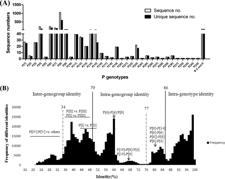
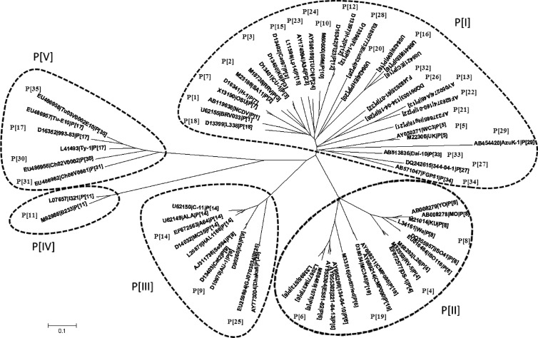
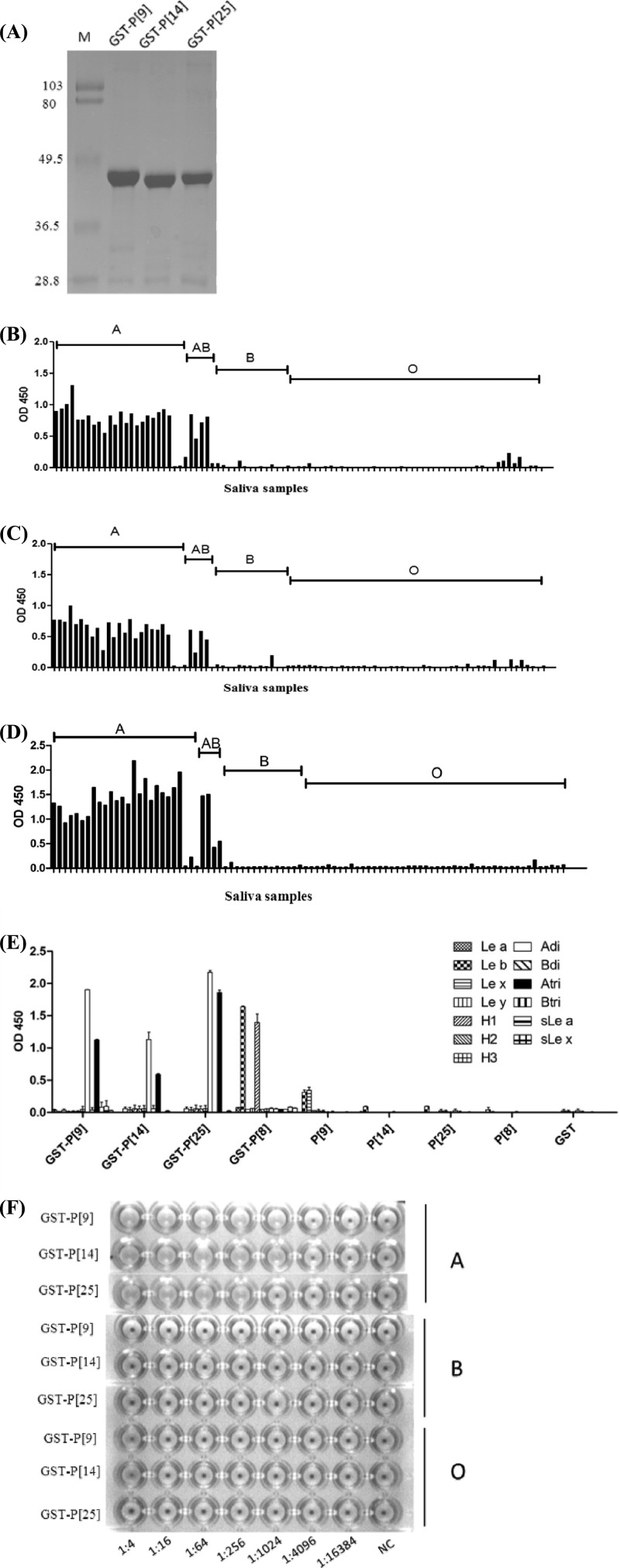
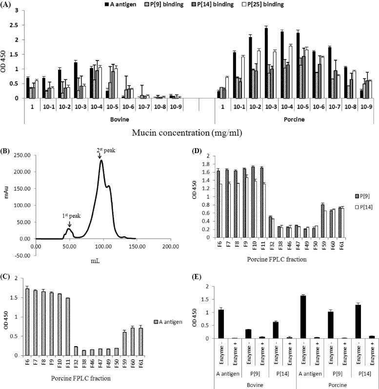
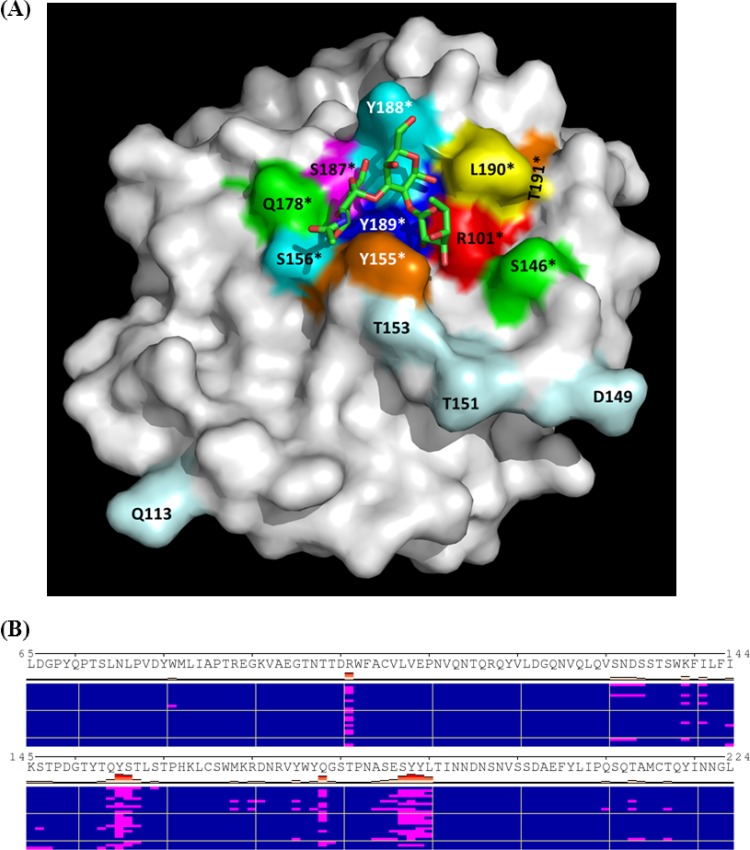
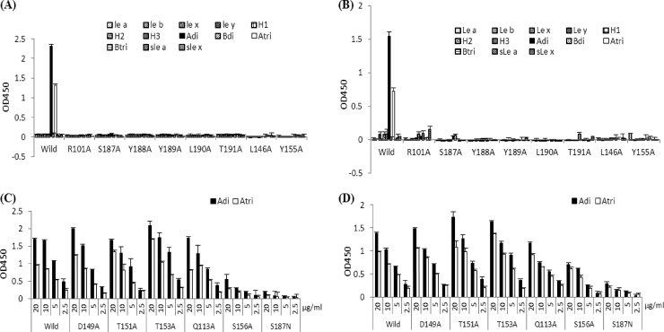
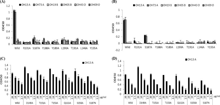
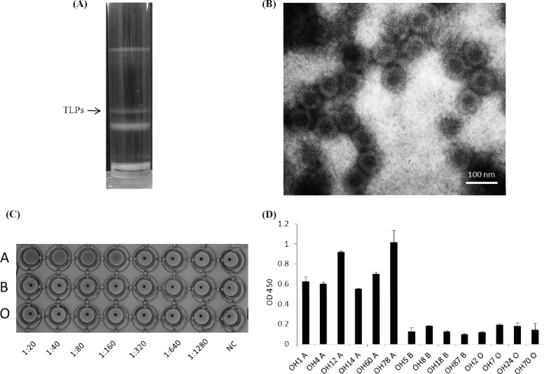
The distal portion of rotavirus (RV) VP4 spike protein (VP8*) is implicated in binding to cellular receptors, thereby facilitating viral attachment and entry. While VP8* of some animal RVs engage sialic acid, human RVs often attach to and enter cells in a sialic acid-independent manner. A recent study demonstrated that the major human RVs (P[4], P[6], and P[8]) recognize human histo-blood group antigens (HBGAs). In this study, we performed a phylogenetic analysis of RVs and showed further variations of RV interaction with HBGAs. On the basis of the VP8* sequences, RVs are grouped into five P genogroups (P[I] to P[V]), of which P[I], P[IV], and P[V] mainly infect animals, P[II] infects humans, and P[III] infects both animals and humans. The sialic acid-dependent RVs (P[1], P[2], P[3], and P[7]) form a subcluster within P[I], while all three major P genotypes of human RVs (P[4], P[6], and P[8]) are clustered in P[II]. We then characterized three human RVs (P[9], P[14], and P[25]) in P[III] and observed a new pattern of binding to the type A antigen which is distinct from that of the P[II] RVs. The binding was demonstrated by hemagglutination and saliva binding assay using recombinant VP8* and native RVs. Homology modeling and mutagenesis study showed that the locations of the carbohydrate binding interfaces are shared with the sialic acid-dependent RVs, although different amino acids are involved. The P[III] VP8* proteins also bind the A antigens of the porcine and bovine mucins, suggesting the A antigen as a possible factor for cross-species transmission of RVs. Our study suggests that HBGAs play an important role in RV infection and evolution.


Figures

Fig 1
Fig 1
Sequence numbers for different P genotypes and the distribution of pairwise sequence identity of rotavirus VP8* proteins. (A) Sequence numbers for each of the genotypes. A total of 2,107 available VP8* sequences in GenBank were grouped into 35 genotypes of group A RVs and group B, C, and D RVs. The unique sequences were also included in this figure after excluding identical sequences concerning the region from aa 46 to 231. Identical sequences in the original VP8* sequence pool were excluded using BioEdit software (version 7.0.9.0). (B) The remaining 1,319 unique group A RV sequences were analyzed for pairwise sequence identities within or between genotypes. The graph was constructed by plotting all the calculated pairwise identities, with the percent identities in the abscissa (x axis) and the frequency of each calculated pairwise identities in the ordinate (y axis). To determine a cutoff value, we chose a ratio below and closest to 1 between inter- and intragenotype identities. Based on this principle, the proposed intra- and intergenotype and intra- and intergenogroup identity ranges for the potential cutoff value are shown. A cutoff value of 86% sequence identity for intergenotypes and a 50% cutoff value for intercluster (genogroup) distance were selected.
Fig 2
Fig 2
Phylogenetic tree of rotavirus VP8*. The phylogenetic tree was generated based on the VP8*sequences (aa 46 to 231) using the neighbor-joining method. Seventy-one prototype and/or reference rotaviruses, representing all 35 known genotypes, were selected. Sequence information for the 71 selected strains is shown in the order of GenBank accession number, strain name, and P genotype.
Fig 3
Fig 3
Hemagglutination and saliva- and synthetic oligosaccharide-based binding of P[9], P[14], and P[25] VP8* proteins. (A) SDS-PAGE analysis of the purified recombinant VP8* proteins of P[9], P[14], and P[25] rotaviruses. The GST-VP8* fusion protein is ∼45 kDa. “M” indicates a prestained protein marker (low range; Bio-Rad), with bands from the top to the bottom representing 103, 80, 49.5, 36.5, and 28.8 kDa, respectively. (B to D) Binding of P[9] (B), P[14] (C), and P[25] (D) GST-VP8* fusion proteins to a panel of well-characterized saliva samples. (E) Binding of the recombinant VP8*proteins to the synthetic oligosaccharides. Polyacrylamide (PAA)-biotin conjugates of Lewis antigens (Lea, Leb, Lex, and Ley), H type1 (H1), H type 2 (H2), H type 3(H3), type A and type B disaccharides (Adi and Bdi), type A and type B trisaccharides (Atri and Btri), sialyl-Lea (sLe a), and sialyl-Lex (sLe x) were used in the binding assay. The GST-VP8* fusion protein from a P[8] strain expressed previously in our lab was also used in the binding assay as a control to confirm the binding specificity observed in this study on the recombinant proteins of P[9], P[14], and P[25]. Recombinant proteins with or without the GST tag and the GST tag alone were tested. (F) Hemagglutination assay of the recombinant VP8* proteins. GST-VP8* proteins were serially diluted with PBS, and the PBS buffer without recombinant proteins was used as the negative control (NC). OD 450, optical density at 450 nm.
Fig 4
Fig 4
Binding of recombinant rotavirus VP8* to porcine and bovine mucins. (A) GST-VP8* fusion proteins of P[9], P[14], and P[25] were tested for binding to serial dilutions of boiled porcine and bovine mucins. The A antigen signal in each dilution is shown. (B) Gel filtration profiles of the porcine mucins. Fractions from each major peak were collected. (C to D) The A antigen signals (C) and the binding of P[9] and P[14] VP8* (D) to these fractions were determined by enzyme-linked immunosorbent assay as shown (F6 to F11 were fractions from the 1st peak, F32 to F50 were fractions from the 2nd peak, and F59 to F61 were fractions after the 2nd peak). (E) Effect of enzymatic removal of the A epitope on the rotavirus VP8* binding to porcine and bovine mucins. F6 from the high-molecular-weight peaks (the 1st peaks) of the porcine and bovine mucins was treated with an N-acetylgalactosaminidase. The signals of the A antigen and the binding of the P[9] and P[14] VP8* to the mucins before and after the enzyme treatment were measured. Error bars show the standard deviations from two experiments. OD 450, optical density at 450 nm.
Fig 5
Fig 5
Modeling of HBGA-binding interface of a P[9] VP8*. The homology model of a P[9] VP8* from ModBase was used for modeling the HBGA binding site by docking simulations. (A) P[9] VP8* is shown in surface representation, with indication of a predicted HBGA-binding pocket that is formed by residues R101, Y155, S156, Q178, S187, Y188, Y189, and L190. The best pose of the A trisaccharide in stick representations with carbon (green), oxygen (red), and nitrogen (blue) atoms of the trisaccharide is shown. The binding mode of the A trisaccharide in the binding pocket was predicted based on a calculated lowest energy. The amino acid residues involved in the HBGA interactions are labeled in color and with asterisks. Amino acid residues away from the binding pocket tested in the mutagenesis study are also labeled in light blue. (B) Clusters of docking poses of the A trisaccharide to VP8* by AutoDock. A total of 23 distinct clusters (of docking poses), each represented by a row, are shown. Residues in contact with the ligand in each pose (highlighted as red bars) are shown in magenta. Frequent hits of the protein by the A trisaccharide at residues around the binding pocket shown in panel A (R101, Y155, S156, Q178, S187, Y188, Y189, and L190) are apparent.
Fig 6
Fig 6
Oligosaccharide binding of VP8* mutants with single amino acid changes in the binding pocket or away from the pocket. Most of the VP8* mutants with a single mutation in the binding pockets lost the binding activities completely for all tested HBGAs for both P[9] (A) and P[14] (B) rotavirus strains (protein concentrations of 10 μg/ml were used in the binding assay). Abbreviations of the oligosaccharide conjugates are the same as in Fig. 3. Mutants that retained binding to the A antigen were further tested for their binding to A di- and trisaccharides with serial dilutions of VP8* proteins in 20, 10, 5, and 2.5 μg/ml, as shown in panels C and D for P[9] and P[14], respectively. Error bars show the standard deviations from three experiments.
Fig 7
Fig 7
Saliva-based binding results of various mutant VP8* proteins with single amino acid changes in or away from the binding interface. Saliva samples representing type A, B, and O secretors were used. Mutants (10 μg/ml) that dramatically lost the ability to bind the type A saliva are shown in panels A and B for P[9] and P[14], respectively. Mutants that retained the ability to bind to type A saliva are shown in panels C and D for P[9] and P[14], respectively, with variable concentrations of the VP8* proteins (20, 10, 5, and 2.5 μg/ml). Error bars show the standard deviations from three experiments.
Fig 8
Fig 8
Purification, hemagglutination (HA), and binding to saliva samples of the triple-layered particles (TLPs) of a P [9] rotavirus (Arg 720 strain). (A) CsCl gradient purification with indication of the TLP band. (B) The TLP band (density, 1.36 g/cm3 in CsCl) was confirmed by electron microscopy (EM) examination. (C) HA of the TLP virions. Twofold serial dilutions of TLPs were used in the HA assay, with PBS buffer without TLPs as the negative control (NC). The TLPs were found to specifically agglutinate the type A RBCs. (D) Specific binding of the TLPs to the type A saliva samples. The x axes show a panel of A-positive and -negative saliva samples, and the y axes show the optical density (OD) value of the binding signals. Error bars show the standard deviations from two experiments.

References

    1. Beards G, Graham C. 1995. Temporal distribution of rotavirus G-serotypes in the West Midlands region of the United Kingdom, 1983–1994. J. Diarrhoeal Dis. Res. 13:235–237 - PubMed
    1. Beards GM, Desselberger U, Flewett TH. 1989. Temporal and geographical distributions of human rotavirus serotypes, 1983 to 1988. J. Clin. Microbiol. 27:2827–2833 - PMC - PubMed
    1. Bishop JR, Gagneux P. 2007. Evolution of carbohydrate antigens—microbial forces shaping host glycomes? Glycobiology 17:23R–34R - PubMed
    1. Bishop RF, Unicomb LE, Barnes GL. 1991. Epidemiology of rotavirus serotypes in Melbourne, Australia, from 1973 to 1989. J. Clin. Microbiol. 29:862–868 - PMC - PubMed
    1. Castello AA, et al. 2009. Characterization of genotype P[9]G12 rotavirus strains from Argentina: high similarity with Japanese and Korean G12 strains J. Med. Virol. 81:371–381 - PubMed

Publication types

MeSH terms

LinkOut - more resources