. 2012 Sep;86(18):9983-94.
doi: 10.1128/JVI.00839-12. Epub 2012 Jul 3.

Kaposi's sarcoma-associated herpesvirus-encoded LANA recruits topoisomerase IIβ for latent DNA replication of the terminal repeats

Affiliations

Kaposi's sarcoma-associated herpesvirus-encoded LANA recruits topoisomerase IIβ for latent DNA replication of the terminal repeats

Pravinkumar Purushothaman et al. J Virol. 2012 Sep.

Abstract

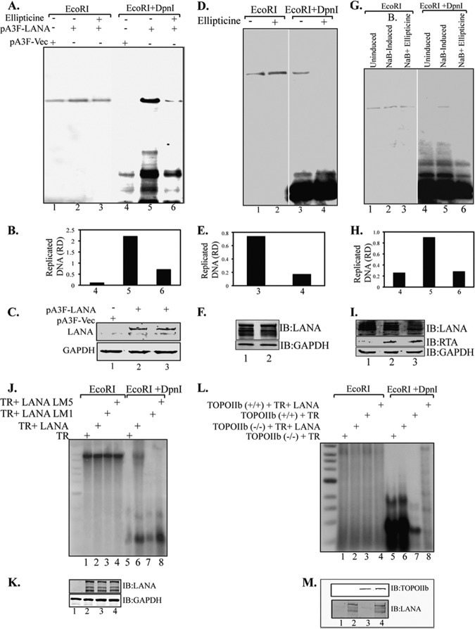
The latency-associated nuclear antigen (LANA) encoded by Kaposi's sarcoma-associated herpesvirus (KSHV) plays a major role in maintaining latency and is critical for the perpetual segregation of viral episomes to the progeny nuclei of newly divided cells. LANA binds to KSHV terminal repeat (TR) DNA and tethers the viral episomes to host chromosomes through the association of chromatin-bound cellular proteins. TR elements serve as potential origin sites of KSHV replication and have been shown to play important roles in latent DNA replication and transcription of adjacent genes. Affinity chromatography and proteomics analysis using KSHV TR DNA and the LANA binding site as the affinity column identified topoisomerase IIβ (TopoIIβ) as a LANA-interacting protein. Here, we show that TopoIIβ forms complexes with LANA that colocalize as punctuate bodies in the nucleus of KSHV-infected cells. The specific TopoIIβ binding region of LANA has been identified to its N terminus and the first 32 amino acid residues containing the nucleosome-binding region crucial for binding. Moreover, this region could also act as a dominant negative to disrupt association of TopoIIβ with LANA. TopoIIβ plays an important role in LANA-dependent latent DNA replication, as addition of ellipticine, a selective inhibitor of TopoII, negatively regulated replication mediated by the TR. DNA break labeling and chromatin immunoprecipitation assay using biotin-16-dUTP and terminal deoxynucleotide transferase showed that TopoIIβ mediates a transient DNA break on viral DNA. These studies confirm that LANA recruits TopoIIβ at the origins of latent replication to unwind the DNA for replication.


Figures

Fig 1
Fig 1
KSHV LANA associates with TopoIIβ in KSHV-positive cells. (A to D) Coimmunoprecipitation assays were performed using 25 million KSHV-positive cells (BCBL-1) with anti-LANA antibody (A); using 25 million BCBL-1 cells with anti-TopoIIβ antibody and subsequent detection with anti-TopoIIβ and LANA antibodies (lane 4) (B); using 25 million KSHV-positive cells (JSC-1) with anti-LANA antibody (C); using 25 million JSC-1 cells with anti-TopoIIβ antibody and subsequent detection with anti-TopoIIβ and LANA antibodies (lane 4) (D). (E) Co-IP analysis with anti-FLAG antibody from 25 million BJAB cells expressing either YFP-Flag (Y-Flag) or LANA-YFP-Flag (Y-LFlag) and subsequent detection with anti-TopoIIβ antibody. TopoIIβ was found to coimmunoprecipitate with exogenously supplied LANA (lane 4).
Fig 2
Fig 2
LANA colocalizes with TopoIIβ in KSHV-positive cells. KSHV-positive cells BCBL-1 and JSC-1 (A) and Bac36 Vero cells (B) were stained with rat anti-LANA and rabbit anti-TopoIIβ antibodies. LANA is shown in green and TopoIIβ in red. Nuclear stain TO-PRO 3 is shown in blue. LANA and TopoIIβ colocalize in the nucleus as punctate bodies. DIC images were used to show the cells' morphology.
Fig 3
Fig 3
The amino terminus of LANA interacts with TopoIIβ. (A) Twenty million HEK 293T cells were transfected with flag epitope-tagged pA3F empty vector, pA3F LANA, pA3F LANA-N, and pA3F LANA-C along with GFP-TopoIIβ. At 36 h posttransfection, cells were harvested and immunoprecipitated with anti-Flag antibody and subsequently detected with anti-TopoIIβ antibody (lanes 6 and 7). (B) Similarly, HEK 293T cells were transfected with pA3F empty, pA3F LANA-N, and pA3F LANA-C along with GFP-TopoIIβ. At 36 h posttransfection, cells were harvested and immunoprecipitated with anti-Flag antibody and subsequently detected with anti-TopoIIβ antibody. TopoIIβ specifically interacts with full-length LANA and the N-terminal region of LANA (panel A, lane 7, and panel B, lane 5). (C) HEK 293T cells were transfected with pA3F empty, pA3F LANA, pA3F LANA-N, LANA 1 to 32, and pA3F LANA-C. At 36 h posttransfection, cells were harvested and immunoprecipitated with anti-Flag antibody and subsequently detected with anti-TopoIIβ and LANA antibodies. Endogenous TopoIIβ specifically interacts with full-length LANA and the N-terminal region of LANA (lanes 7, 8, and 9). (D) HEK 293T cells were transfected with pA3F empty, pA3F LANA, and pA3F LANA-N along with GFP-TopoIIβ. At 36 h posttransfection, cells were harvested and the lysate was incubated with 2,000 U micrococcal nuclease (NEB) for 30 min at 37°C and immunoprecipitated with anti-Flag antibody and subsequently detected with anti-TopoIIβ antibody. TopoIIβ interacts specifically with full-length LANA and its amino terminus irrespective of nuclease treatment (lanes 5 and 6). (E) Micrococcal nuclease-digested DNA on ethidium bromide (EtBr)-stained agarose gel.
Fig 4
Fig 4
The aa 1 to 32 region at the amino terminus of LANA is responsible for the association with TopoIIβ. (A) Schematic showing the truncations of LANA N-terminal region. (B) Twenty million HEK 293T cells were transfected with pEGP-Myc LANA aa 33 to 275, pEGFP-Myc LANA aa 1 to 250, pEGFP-Myc LANA aa 1 to 150, and pEGFP-Myc LANA aa 33 to 340 with GFP-TopoIIβ. At 36 h posttransfection, cells were harvested and immunoprecipitated with anti-Myc antibody (9E10) and subsequently detected with anti-TopoIIβ antibody (lanes 6 and 7). (C) Twenty-five million BJAB cells stably expressing pEGP-Myc empty, pEGP-Myc LANA aa 1 to 32, and pEGP-Myc LANA aa 1 to 32 with substitutions at aa 5 to 15 (5aa15) were harvested and immunoprecipitated with anti-Myc antibody (9E10) and subsequently detected with anti-TopoIIβ antibody (lane 5). (D) HEK 293 T cells were transfected with pEGFP-Myc empty, pEGFP-Myc LANA aa 1 to 32, pEGFP-Myc LANA aa 1 to 32 5aa15, pEGFP-Myc LANA aa 1 to 340, and pEGFP-Myc LANA aa 1 to 340 5aa15 with GFP-TopoIIβ. At 36 h posttransfection, cells were harvested and immunoprecipitated with anti-Myc antibody (9E10) and subsequently detected with anti-TopoIIβ antibody (lanes 7 and 9). (E) In vitro GST binding: GST, GST-LANA aa 1 to 32, and GST-LANA aa 1 to 32 5aa15 fusion proteins were expressed in E. coli, purified with glutathione-Sepharose beads, and incubated with TopoIIβ cell lysate prepared from HEK 293T cells transfected with GFP-TopoIIβ. The aa 1 to 32 region of LANA interacted with TopoIIβ (lane 2).
Fig 5
Fig 5
The aa 1 to 32 region of the amino terminus of LANA is responsible for TopoIIβ association. (A) Schematic showing the alanine substitution mutations of aa 1 to 32 of the LANA N-terminal region. (B) Twenty million HEK 293T cells were transfected with pEGFP-Myc empty, pEGFP-Myc LANA aa 1 to 32, pEGFP-Myc LANA aa 1 to 340, pEGFP-Myc LANA aa 1 to 340 M1, pEGFP-Myc LANA aa 1 to 340 M2, pEGFP-Myc LANA aa 1 to 340 M3, pEGFP-Myc LANA aa 1 to 340 M4, and pEGFP-Myc LANA aa 1 to 340 M5 with GFP-TopoIIβ. At 36 h posttransfection, cells were harvested and immunoprecipitated with anti-Myc antibody (9E10) and subsequently detected with anti-TopoIIβ antibody. (C) Similarly, HEK 293T cells were transfected with pEGFP-Myc empty, pEGFP-Myc LANA aa 1 to 32, pEGFP-Myc LANA aa 1 to 340, pEGFP-Myc LANA aa 1 to 32 M1, pEGFP-Myc LANA aa 1 to 32 M2, pEGFP-Myc LANA aa 1 to 32 M3, pEGFP-Myc LANA aa 1 to 32 M4, pEGFP-Myc LANA aa 1 to 32 M5 with GFP-TopoIIβ. At 36 h posttransfection, cells were harvested and immunoprecipitated with anti-Myc antibody (9E10) and subsequently detected with anti-TopoIIβ antibody. The residues 8 to 15 of LANA are crucial for binding TopoIIβ (B and C).
Fig 6
Fig 6
TopoIIβ is required for KSHV latent DNA replication. (A) HEK 293L cells were transfected with TR plasmid along with pA3F-LANA or empty vector pA3F. Cells were treated with ellipticine 24 h posttransfection. Ninety-six hours posttransfection, cells were harvested, and the DNA extracted by Hirt's procedure was subjected to Southern blotting with a TR probe after digestion. Cells expressing LANA without ellipticine treatment show a prominent DpnI-resistant replicated DNA (lane 5), whereas cells treated with ellipticine showed a faint band (lane 6). (B) Quantitation of the replicated DNA based on the relative density (RD). (C) Expression of LANA and GAPDH. (D) BCBL-1 cells transfected with TR plasmid were treated with ellipticine 24 h posttransfection. Ninety-six hours posttransfection, cells were harvested, and the DNA extracted by Hirt's procedure was subjected to Southern blotting with TR probe after digestion. Ellipticine effectively blocked KSHV latent DNA replication in KSHV-positive cells (lane 4). (E) Quantitation of the replicated DNA. (F) Expression levels of LANA and GAPDH. (G) BCBL-1 cells transfected with ori-Lyt plasmid were first induced with 3 mM sodium butyrate and 20 μg/ml TPA followed by treatment with ellipticine 24 h posttransfection. Ninety-six hours posttransfection, cells were harvested, and the DNA extracted by Hirt's procedure was subjected to Southern blotting with ori-Lyt probe after digestion. Ellipticine blocked KSHV lytic DNA replication (lane 6). (H) Quantitation of the replicated DNA. (I) Western blots showing the expression of LANA, RTA, and GAPDH. (J) LANA mutants M1 and M5 do not support KSHV latent DNA replication (lanes 7 and 8): HEK 293L cells were transfected with TR plasmid along with pA3M-LANA, pA3M-LANA-M1, or pA3M-LANA-M5. Ninety-six hours posttransfection, cells were harvested, and the DNA extracted by Hirt's procedure was subjected to Southern blotting with TR probe after digestion. (K) Expression of LANA and GAPDH. (L) TopoIIβ is required for KSHV latent DNA replication: wild-type (TopoIIβ+/+) and TopoIIβ knockout (TopoIIβ−/−) MEF NIH 3T3 cells were transfected with TR plasmid along with empty vector pA3F or pA3F-LANA. Ninety-six hours posttransfection, cells were harvested, and the DNA extracted by Hirt's procedure was subjected to Southern blotting with TR probe after digestion. (M) Expression of LANA and TopoIIβ in the cells used for replication assays.
Fig 7
Fig 7
The minimal binding region of LANA acts as a dominant negative to disrupt LANA-TopoIIβ interaction and replication. (A) Twenty million HEK 293T cells were transfected with GFP-TopoIIβ along with pA3F, pA3F-LANA, pA3F-LANA C terminus, pA3F-LANA N, and pA3F-LANA N with two different amounts (20 μg and 40 μg) of GFP-Myc-LANA aa 1 to 32 and 40 μg GFP-Myc-LANA aa 1 to 32 mutant M5 (with alanine substitutions at aa 5 to 15). At 36 h posttransfection, cells were harvested and immunoprecipitated with anti-flag antibody to precipitate LANA and its mutants, followed by detection of TopoIIβ as the coimmunoprecipitated proteins. LANA aa 1 to 32 was found to interfere with LANA TopoIIβ interaction (lanes 5 and 6), whereas transfection of LANA aa 1 to 32 mutant M5 did not interfere with LANA and TopoIIβ interaction (lane 7). Expression of increased amounts of LANA aa 1 to 32 further decreased TopoIIβ binding (lane 6). Expressions of LANA aa 1 to 32 were detected by anti-Myc immunoblot (asterisk). (B) HEK 293L cells were transfected with TR plasmid along with GFP-Myc-LANA 1 to 32, pA3F-LANA, or pA3F-LANA with two different amounts (20 and 40 μg) of GFP-Myc-LANA 1 to 32 and pA3F-LANA along with 40 μg of GFP-Myc-LANA 1 to 32 M5. Ninety-six hours posttransfection, cells were harvested, and the DNA extracted by Hirt's procedure was subjected to Southern blotting with TR probe after digestion. LANA aa 1 to 32 interfere with KSHV latent DNA replication as detected by a reduced level of DpnI-resistant band (compare lanes 9 and 10). Increasing expression of LANA aa 1 to 32 further reduced the replication of TR plasmid (compare lanes 11 and 10), however; mutant 5 (M5) of LANA aa 1 to 32 was unable to suppress replication (compare lanes 11 and 12). (C) Quantitation of the replicated DNA. (D) Expression levels of LANA and GFP-Myc-LANA aa 1 to 32 and GFP-Myc-LANA aa 1 to 32 M5. (E) Schematic showing DNA break labeling and ChIP assay procedure. Wild-type (TopoIIβ+/+) and TopoIIβ knockdown (TopoIIβ−/−) MEF NIH 3T3 cells were transfected with KSHV TR plasmid either with pA3F-LANA or with empty vector pA3F. The nuclei were subsequently labeled with biotin-16-dUTP using terminal deoxynucleotidyl transferase (TdT) and subjected to chromatin immunoprecipitation (ChIP). (F) Quantitative real-time PCR was done on the input and ChIP DNA samples using KSHV TR-specific primers. Relative copies of the dUTP-labeled ChIP DNA were determined as the ratio of cells without LANA to cells with LANA in TopoIIβ−/− and TopoIIβ+/+ cells to determine the effect of LANA on dsDNA breaks. TopoIIβ+/+ cells showed significant increase in the presence of LANA. (G) DNA break labeling and ChIP assay was performed on HEK 293L cells transfected with KSHV TR-containing plasmids with either pA3F-LANA or the empty vector pA3F. One set of cells with LANA with TR was treated with 5 μM ellipticine. dUTP-labeled DNAs were determined in a real-time PCR using primer for TR (black bar) and vector backbone (gray bar). An increase in dUTP-labeled ChIP DNA in the TR region but not in the ampicillin region (vector) in LANA-expressing cells suggests the existence of a dsDNA break in the TR region. Ellipticine treatment blocked dUTP incorporation, suggesting a TopoII-mediated dsDNA break. (H) At 24 h posttransfection, 293L cells were harvested for flow cytometry and cell cycle analysis.
Fig 8
Fig 8
Schematic model showing the association of LANA with TopoIIβ. LANA recruits TopoIIβ to the sites of latent origin of replication. TopoIIβ in turn mediates double-stranded breaks required for viral DNA replication, further facilitated by the cellular replication machinery.

Similar articles

Cited by

References

    1. An FQ, et al. 2005. The latency-associated nuclear antigen of Kaposi's sarcoma-associated herpesvirus modulates cellular gene expression and protects lymphoid cells from p16 INK4A-induced cell cycle arrest. J. Biol. Chem. 280:3862–3874 - PubMed
    1. Ballestas ME, Chatis PA, Kaye KM. 1999. Efficient persistence of extrachromosomal KSHV DNA mediated by latency-associated nuclear antigen. Science 284:641–644 - PubMed
    1. Ballestas ME, Kaye KM. 2001. Kaposi's sarcoma-associated herpesvirus latency-associated nuclear antigen 1 mediates episome persistence through cis-acting terminal repeat (TR) sequence and specifically binds TR DNA. J. Virol. 75:3250–3258 - PMC - PubMed
    1. Ballestas ME, Kaye KM. 2011. The latency-associated nuclear antigen, a multifunctional protein central to Kaposi's sarcoma-associated herpesvirus latency. Future Microbiol. 6:1399–1413 - PMC - PubMed
    1. Barbera AJ, Ballestas ME, Kaye KM. 2004. The Kaposi's sarcoma-associated herpesvirus latency-associated nuclear antigen 1 N terminus is essential for chromosome association, DNA replication, and episome persistence. J. Virol. 78:294–301 - PMC - PubMed

Publication types

MeSH terms

LinkOut - more resources