. 2012 Sep;86(18):9929-40.
doi: 10.1128/JVI.01154-12. Epub 2012 Jul 3.

Virion-associated complement regulator CD55 is more potent than CD46 in mediating resistance of mumps virus and vesicular stomatitis virus to neutralization

Affiliations

Virion-associated complement regulator CD55 is more potent than CD46 in mediating resistance of mumps virus and vesicular stomatitis virus to neutralization

John B Johnson et al. J Virol. 2012 Sep.

Abstract

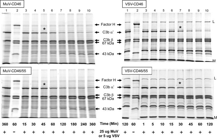
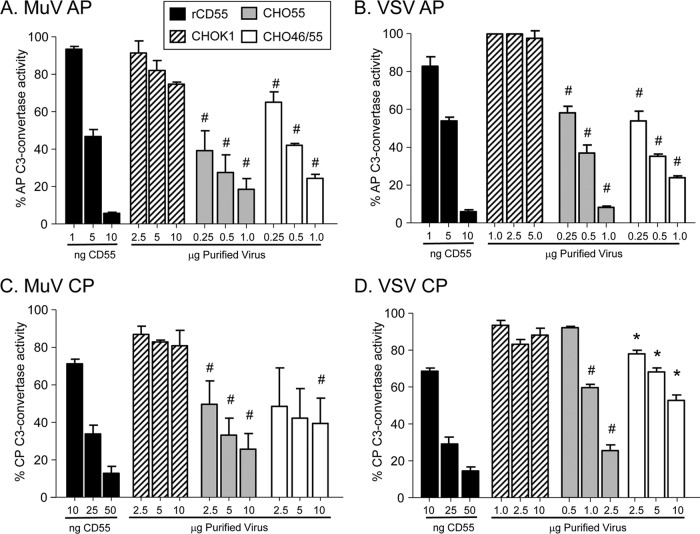
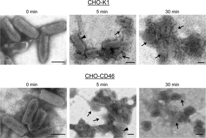
Enveloped viruses can incorporate host cell membrane proteins during the budding process. Here we demonstrate that mumps virus (MuV) and vesicular stomatitis virus (VSV) assemble to include CD46 and CD55, two host cell regulators which inhibit propagation of complement pathways through distinct mechanisms. Using viruses which incorporated CD46 alone, CD55 alone, or both CD46 and CD55, we have tested the relative contribution of these regulators in resistance to complement-mediated neutralization. Virion-associated CD46 and CD55 were biologically active, with VSV showing higher levels of activity of both cofactors, which promoted factor I-mediated cleavage of C3b into iC3b as well as decay-accelerating factor (DAF) activity against the C3 convertase, than MuV. Time courses of in vitro neutralization with normal human serum (NHS) showed that both regulators could delay neutralization, but viruses containing CD46 alone were neutralized faster and more completely than viruses containing CD55 alone. A dominant inhibitory role for CD55 was most evident for VSV, where virus containing CD55 alone was not substantially different in neutralization kinetics from virus harboring both regulators. Electron microscopy showed that VSV neutralization proceeded through virion aggregation followed by lysis, with virion-associated CD55 providing a delay in both aggregation and lysis more substantial than that conferred by CD46. Our results demonstrate the functional significance of incorporation of host cell factors during virion envelope assembly. They also define pathways of virus complement-mediated neutralization and suggest the design of more effective viral vectors.


Figures

Fig 1
Fig 1
NHS-mediated neutralization of HeLa-grown MuV is delayed compared to that of CHO-grown MuV. (A) MuV was grown in HeLa or CHO cells and purified by gradient centrifugation. Virions were analyzed for levels of CD55, CD46, and P protein by Western blotting. (B) One hundred PFU of virus was incubated with NHS for the indicated times, and the remaining infectivity was determined by plaque assay. No loss of infectivity was seen using HI NHS.
Fig 2
Fig 2
CD55 and CD46 expression in CHO cell lines. (A) Western blotting. Lysates from control CHO-K1, CHO-CD55, CHO-CD46, or double-positive CHO-CD46/55 cells were analyzed by Western blotting for expression of CD46, CD55, and actin. (B) Cell surface expression of CD46 and CD55. CHO cells expressing CD46, CD55, or both regulators were analyzed by flow cytometry for cell surface expression of CD46 and CD55. MFIs (arbitrary units on y axis; number of cells on x axis) are shown in parentheses.
Fig 3
Fig 3
CD46 and CD55 are associated with purified MuV virions. (A) Western blotting. MuV derived from the indicated cell lines was purified by gradient centrifugation and analyzed by Western blotting for the presence of CD46 or CD55. Levels of viral P protein were analyzed as a loading control. (B) EM analysis. Purified virus derived from CHO cells expressing CD46 (left), CD55 (middle), or both CD46 and CD55 (right) was treated with the indicated antibodies, followed by 6-nm (CD46) or 12-nm (CD55) colloidal gold-labeled goat anti-mouse antibody (top row). Control samples were treated with secondary antibody alone (bottom row). Samples were analyzed by EM at a magnification of ×55,000. Bars, 0.1 μm. White and open arrows, locations of staining for CD46 and CD55, respectively; 1° and 2°, primary and secondary antibodies, respectively.
Fig 4
Fig 4
CD46 and CD55 are associated with purified VSV virions. (A) Western blotting. Purified VSV derived from the indicated cell lines was analyzed by Western blotting for the presence of CD46 or CD55. Levels of viral M protein were analyzed as a loading control. (B) EM analysis. Purified virus derived from CHO cells expressing CD46 (left), CD55 (middle), or both CD46 and CD55 (right) was treated with the indicated antibodies, followed by 6-nm (CD46) or 12-nm (CD55) colloidal gold-labeled goat anti-mouse antibody (top row). Control samples were treated with secondary antibody alone (bottom row). Samples were analyzed by EM at a magnification of ×55,000. Bars, 0.1 μm. White and open arrows, locations of staining for CD46 and CD55, respectively.
Fig 5
Fig 5
Cofactor activity associated with MuV and VSV grown in CD46 and CD46/55 CHO cells. Twenty-five micrograms or 5 μg of purified MuV and VSV derived from CHO-CD46 or CHO-CD46/55 cells was incubated for the indicated times with purified C3b and factor I as detailed in Materials and Methods. The cleavage of the alpha′ chain of C3b into the 67- and 43-kDa products was monitored by SDS-PAGE and Coomassie blue staining. Lanes 1 and 2, samples showing negative-control starting products and positive-control products generated by cleavage of C3b-alpha′ by factor I and factor H. The positions of factor H, alpha′, and beta chains of C3b and the 67- and 43-kDa cleavage products are indicated. *, approximate time point when half of C3b-alpha′ was cleaved.
Fig 6
Fig 6
Alternative and classical pathway DAF activity associated with MuV and VSV particles. Purified MuV (A and C) or VSV (B and D) that was derived from the indicated cells was assayed for decay-accelerating activity against the alternative pathway (AP) (A and B) or classical pathway (CP) (C and D) C3 convertase as detailed in Materials and Methods. C3-mediated hemolytic activity is expressed as a percentage of the lysed control sample set at 100%. Data are from triplicate samples, and error bars represent standard deviations. (A and B) #, P < 0.001 when comparing results for 0.25 to 1 μg of MuV-CD55 and MuV-CD46/55 with those for 0.25 to 10 μg from control virus derived from CHO-K1 cells or 0.25 to 1 μg of VSV-CD55 and VSV-CD46/55 with those for 1 to 5 μg from control virus derived from CHO-K1 cells; (C and D) #, P < 0.001 when comparing results for 0.5 to 2.5 μg VSV-CD55 with those for 1 to 10 μg control virus and comparing results for 2.5 to 10 μg MuV-CD55 and MuV-CD46/55 with those for the corresponding amount (μg) of control virus; *, P < 0.01 when comparing results for 0.25 to 10 μg VSV-CD46/55 with those for 1 to 10 μg control virus derived from CHO-K1 cells.
Fig 7
Fig 7
Rate of in vitro neutralization of CD46- and CD55-containing MuV and VSV. One hundred PFU of MuV (A) or VSV (B) derived from the indicated cell lines was incubated with NHS from two different donors for the indicated times, and then remaining infectivity was determined by plaque assay. As a control, virus was incubated with PBS alone for 1 h. Data are from six independent reactions, and error bars represent standard deviations. ^, P < 0.0001 when comparing virus with CD46 only to virus from control CHO-K1 cells; *, P < 0.0001 when comparing virus containing CD55 only to virus containing CD46 only.
Fig 8
Fig 8
Effect of NHS dilution and HI NHS on neutralization of various CHO-grown viruses. (A) Effect of NHS dilution. One hundred PFU of the indicated viruses was incubated for 1 h with the indicated dilutions of NHS, and remaining infectivity was determined by plaque assay. (B) Effect of HI NHS. One hundred PFU of the indicated viruses was incubated for 1 h with PBS, NHS, or HI NHS, and remaining infectivity was determined by plaque assay.
Fig 9
Fig 9
Electron micrographs of in vitro neutralization of VSV derived from CHO-K1 and CHO-CD46 cells. Purified VSV particles derived from the indicated CHO cells were incubated for the indicated times at 37°C with NHS and then analyzed by negative staining and EM. Arrowheads, apparently intact virions; arrows, clearly lysed particles or nucleocapsid-like structures. Bars, 0.1 μm.
Fig 10
Fig 10
Electron micrographs of in vitro neutralization of VSV derived from CHO-CD55 and CHO-CD46/55 cells. Purified VSV particles derived from the indicated CHO cells were incubated for the indicated times at 37°C with NHS and then analyzed by negative staining and EM. Arrowheads, apparently intact virions, arrows, clearly lysed particles or nucleocapsid-like structures. Bars, 0.1 μm.

Similar articles

Cited by

References

    1. Ahmed M, Puckett S, Lyles DS. 2010. Susceptibility of breast cancer cells to an oncolytic matrix (M) protein mutant of vesicular stomatitis virus. Cancer Gene Ther. 17:883–892 - PMC - PubMed
    1. Atkinson JP, Liszewski MK, Richards A, Kavanagh D, Moulton EA. 2005. Hemolytic uremic syndrome: an example of insufficient complement regulation on self tissue. Ann. N. Y. Acad. Sci. 1056:144–152 - PubMed
    1. Beebe DP, Cooper NR. 1981. Neutralization of vesicular stomatitis virus (VSV) by human complement requires a natural IgM antibody present in human serum. J. Immunol. 126:1562–1568 - PubMed
    1. Bernet J, Mullick J, Panse Y, Parab PB, Sahu A. 2004. Kinetic analysis of the interactions between vaccinia virus complement control protein and human complement proteins C3b and C4b. J. Virol. 78:9446–9457 - PMC - PubMed
    1. Blue CE, Spiller OB, Blackbourn DJ. 2004. The relevance of complement to virus biology. Virology 319:176–184 - PMC - PubMed

Publication types

MeSH terms

LinkOut - more resources