. 2012 Sep;86(17):9484-94.
doi: 10.1128/JVI.01114-12. Epub 2012 Jul 3.

Adaptive changes in alphavirus mRNA translation allowed colonization of vertebrate hosts

Affiliations

Adaptive changes in alphavirus mRNA translation allowed colonization of vertebrate hosts

Iván Ventoso. J Virol. 2012 Sep.

Abstract

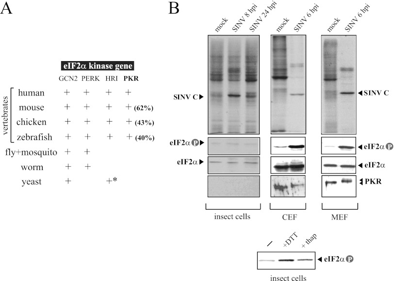
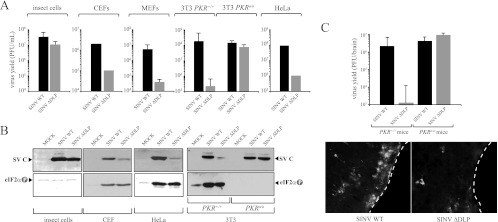
Members of the Alphavirus genus are arboviruses that alternate replication in mosquitoes and vertebrate hosts. In vertebrate cells, the alphavirus resists the activation of antiviral RNA-activated protein kinase (PKR) by the presence of a prominent RNA structure (downstream loop [DLP]) located in viral 26S transcripts, which allows an eIF2-independent translation initiation of these mRNAs. This article shows that DLP structure is essential for replication of Sindbis virus (SINV) in vertebrate cell lines and animals but is dispensable for replication in insect cells, where no ortholog of the vertebrate PKR gene has been found. Sequence comparisons and structural RNA analysis revealed the evolutionary conservation of DLP in SINV and predicted the existence of equivalent DLP structures in many members of the Alphavirus genus. A mutant SINV lacking the DLP structure evolved in murine cells to recover a wild-type phenotype by creating an alternative structure in the RNA that restored the translational independence for eIF2. Genetic, phylogenetic, and biochemical data presented here support an evolutionary scenario for the natural history of alphaviruses, in which the acquisition of DLP structure in their mRNAs probably allowed the colonization of vertebrate host and the consequent geographic expansion of some of these viruses worldwide.


Figures

Fig 1
Fig 1
Differential responses of insect and vertebrate cells to SINV infection. (A) The PKR gene is only present in vertebrates. The result of a BLAST searching for orthologs of eIF2α kinases in different species is summarized in this table. Amino acid sequence identity with human PKR is shown in parentheses. An asterisk denotes the presence of HRI-related genes only in Schizosaccharomyces pombe. (B) Effect of SINV infection on translation, PKR activation and eIF2α phosphorylation in insect (H5), avian (CEF), and murine (MEF) cells. Cells were infected at an MOI of 25 PFU/cell and at the indicated times pulsed with [35S]Met/Cys for 30 min, and labeled proteins were analyzed by autoradiography as described in Materials and Methods (upper panels). Extracts were also analyzed by immunoblotting against phospho-eIF2α, total eIF2α, and PKR. Note that the insect eIF2α band migrated more slowly than the vertebrate eIF2α protein. In parallel experiments, H5 cells were treated for 2 h with 1 mM DTT and 10 μM thapsigargin, two stressors that induced eIF2α phosphorylation by activating the endoplasmic reticulum-associated eIF2α kinase (PERK) (lower panel).
Fig 2
Fig 2
DLP structure in SINV 26S mRNA is essential for virus replication in vertebrate hosts. (A) Viral yields of WT and ΔDLP mutant viruses in insect cells (H5), chicken cells (CEF), murine cells (MEF and 3T3 derived from WT and PKR knockout mice), and human cells (HeLa). For CEF and HeLa cells, the data are the means of two independent experiments. For the rest, the means ± standard deviations (SD) of four independent experiments are shown. (B) Synthesis of capsid protein and eIF2α phosphorylation in the different cell lines infected with WT and ΔDLP mutant viruses. (C) DLP structure is essential for replication of SINV in mice. Wild-type and PKRo/o mice were infected with the indicated virus, and 4 days later, animals were sacrificed and virus yields in whole brains were titrated on BHK21 cells as described in Materials and Methods. This graph is similar to that published previously (14), but now more animals per group (n = 7) are included. Data are expressed as means ± SD. Brains of some infected animals were cryosectioned and analyzed by immunofluorescence (IF) using an anti-SINV capsid antiserum as described in Materials and Methods. White dashed lines show the external border of cerebral cortex.
Fig 3
Fig 3
Structural features of DLPs in Alphavirus. (A) Two main topologies were found; type A, a large spiral with some unpaired nucleotides, and type B, a compact spiral. In some cases, a second stem-loop is localized just downstream from the first (bipartite structure). The distance (n) from the initiation codon (AUG) to the base of DLP is shown. (B) Features of DLP among representative members of Alphavirus genus, including topology, stability, G+C composition, and n distances. The vertebrate hosts that act as primary or secondary virus reservoir, and the geographic distribution for each virus is shown. (Data were taken from the Arbovirus Division of CDC [DVBID] and from the International Committee of Taxonomy of Viruses [ICTVdB].) No vertebrate host for AURAV has been described to date (−). MFE, minimum free energy. (C) DLP sequence and structure are conserved among subspecies, genotypes, and isolates of SINV worldwide (see Materials and Methods). Variability along the first 400 nt of 26S mRNA was calculated by the Shannon entropy (H) method. The position of the initiation codon (AUG) is shown as well as the region that encompasses the DLP structure. The lower panel shows the distribution of variants along the first 168 nt of SINV 26S mRNA. The topology of SINV DLP has been confirmed before by enzymatic probing (54). Data were scored as follows: highly variable positions (red) with H > 0.7 affected for >30% of genotypes, moderately variable positions (yellow) with 0.3 < H < 0.7 affected for 10 to 30% of genotypes, and low-variability positions (gray) with H < 0.3 affected for less than 10% of genotypes. Arrowheads show positions where insertion or deletion of few nucleotides in some variants was found (indels). The initiation codon is marked with an arrow.
Fig 4
Fig 4
Some evolutionary patterns of DLPs in Alphavirus. (A) Flexibility of the N-terminal region of the capsid protein to changes allowed the appearance of DLP structures in the alphavirus. The variability in capsid protein sequences among representative alphaviruses was calculated by the Shannon entropy method as described in Materials and Methods. The domain organization of the SINV prototype of the capsid protein is shown. DLP structures are localized in the coding regions of 26S mRNA corresponding to amino acids (aa) 7 to 40 of the capsid protein. (B) Conservation of DLP structure and the corresponding amino acid sequence among species of the SFV clade (SFV, BEBV, UNAV, MIDV, GETV, Sagiyama virus, and RRV). Shown is a phylogenetic tree of the SFV clade based on the first 150 nt of capsid coding sequences that include the DLP structures. Phylogenetic analysis was carried out by the maximum likelihood method using the GTR+I+Γ substitution model. Neighbor-joining (BioNJ), Bayesian (MrBayes), and maximum parsimony (MP) analysis resulted in an almost equivalent tree topology. The numbers shown are the branch support by 1,000 bootstrap resamplings for BioNJ, PhyML, and MP and the posterior probabilities for MrBayes, as indicated in the legend. Branches with <60% support value were collapsed. According to this model, a stable DLP may have emerged at the base of SFV/BEBV/UNAV/MIDV/GETV/Sagiyama virus/RRV clade that further evolved in the RRV/GETV/Sagiyama virus branch (denoted by black dots). The conserved nucleotide framework of DLPs is in bold, whereas the variable positions among viral species of the same clade are denoted by “N.” The variability in capsid protein sequences (Shannon entropy) among members of the SFV/BEBV/UNAV/MIDV/GETV/Sagiyama virus/RRV complex is shown in the lower panel.
Fig 5
Fig 5
ΔDLP mutant virus evolved in murine cells to restore DLP activity. (A) Reversion from the 5th to 6th passages allowed fitness gain and rapid enrichment of cultures with revertant viruses. Viral yields for each passage were titrated. Capsid protein synthesis in NIH 3T3 cells infected with virus preparation from each passage is shown. (B) Revertant virus restored translation of 26S mRNA in NIH 3T3 cells. WT and PKRo/o cells were infected with the reconstituted revertant virus, and the synthesis of capsid protein was compared with that of the WT and parental ΔDLP mutant. Capsid bands were numbered according to the AUG codon from which they are translated. The sizes of the arrows are proportional to the translational efficiency from each AUG. (C) Genotype of revertant virus. The first 350 nt of 26S mRNA are shown. Arrowheads mark the insertion of the 84-nt fragment that resulted from the tandem duplication of the RNA fragment (see the text for details). AUG 1 (#1) is the natural initiation codon AUG50, AUG 2 (#2) is AUG72, and AUG 3 (#3) is AUG107. Note that the revertant has a duplicated no. 3 AUG (termed 3′), which was used as an initiation codon at an extremely low rate. The seven A's labeled in boldface are the mutations introduced in the parental WT virus to destroy the DLP structure. (D) RNA secondary structure of ΔDLP virus. MFE corresponds to nt 49 to 257 of 26S mRNA, although only the structure for the fragment from nt 49 to 173 is shown. Shape reactivity was scored as follows: +++, 50 to 100% of maximal; ++, 25 to 50% of maximal; +, <25% of maximal. Noncolored bases show no reactivity. (E) RNA secondary structure of 49 to 257 nt of revertant virus 26S mRNA. Note the new stem-loop formed at 26 nt downstream of AUG 3. Nucleotides that resulted from the tandem duplication are in bold, and the arrowhead indicates the site of insertion. Shape reactivity was scored as indicated above.
Fig 6
Fig 6
AURAV is an isolated member of the SINV group with a suboptimal DLP structure. (A) Phylogenetic tree showing the evolutionary relationships among species of the SINV group (upper). AURAV is the only New World representative member of this group and the most divergent. Amino acid sequences of entire structural region were used for maximum likelihood (PhyML) analysis with JTT+I+Γ substitution model and SFV sequence as an outgroup. Bootstrap confidence is shown. BioNJ, Bayesian analysis, and MP resulted in identical topology. A black dot denotes the emergence of the functional DLP in the SINV clade. The topology and stability of AURAV DLP compared to those of SINV are shown (lower). The geographic distribution of AURAV and SINV genotypes is also shown (Division of Vector-Borne Diseases, Centers for Disease Control and Prevention). (B) Replication of AURAV in insect and vertebrate cell lines. Cells were infected at an MOI of 5 PFU/cell, and virus yields were determined at 2 and 6 days postinfection (dpi) for insect cells (H5) and at 2 dpi for avian and rodent cells. (C) Mice of the indicated genotype were infected with 107 PFU of SINV and AURAV by the intranasal route, and virus yields in brains at 4 dpi were estimated by plaque assay on BHK21 cells as described in Materials and Methods. No virus replication was detected in other main organs, such as the lung and liver. Data are means ± SD from four mice per group. (D) Protein synthesis and eIF2α phosphorylation in cell lines infected with AURAV at a high multiplicity of infection (25 PFU/cell). The upper panel shows autoradiography of [35S]Met-labeled proteins at 6 hpi. Viral capsid and glycoprotein (E1/E2) bands are indicated. The lower panels show anti-PKR and anti-phospho eIF2α blots of corresponding samples.
Fig 7
Fig 7
An evolutionary scenario for the natural history of Alphavirus is proposed. Assuming a New World origin from an insect-borne ancestor, the acquisition of DLP structures in 26S mRNA allowed the colonization of a new vertebrate host (e.g., migratory birds) that may have spread SINV and SFV to the Old World. Cycles involving mosquitoes and vertebrate hosts at present are schematically drawn. In insects, alphaviral 26S mRNA is translated by the canonical mechanism of initiation imposed by eIF2, whereas in vertebrate hosts, it occurs by an eIF2-independent mechanism that requires the DLP structure.

Similar articles

Cited by

References

    1. Aarti I, Rajesh K, Ramaiah KV. 2010. Phosphorylation of eIF2 alpha in Sf9 cells: a stress, survival and suicidal signal. Apoptosis 15:679–692 - PubMed
    1. Abraham N, et al. 1999. Characterization of transgenic mice with targeted disruption of the catalytic domain of the double-stranded RNA-dependent protein kinase, PKR. J. Biol. Chem. 274:5953–5962 - PubMed
    1. Berlanga JJ, et al. 2006. Antiviral effect of the mammalian translation initiation factor 2alpha kinase GCN2 against RNA viruses. EMBO J. 25:1730–1740 - PMC - PubMed
    1. Binder GK, Griffin DE. 2001. Interferon-gamma-mediated site-specific clearance of alphavirus from CNS neurons. Science 293:303–306 - PubMed
    1. Choi HK, et al. 1991. Structure of Sindbis virus core protein reveals a chymotrypsin-like serine proteinase and the organization of the virion. Nature 354:37–43 - PubMed

Publication types

MeSH terms