. 2012 Oct;40(18):9073-88.
doi: 10.1093/nar/gks424. Epub 2012 Jul 3.

Mammalian NUMT insertion is non-random

Affiliations

Mammalian NUMT insertion is non-random

Junko Tsuji et al. Nucleic Acids Res. 2012 Oct.

Abstract

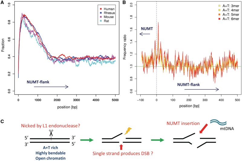
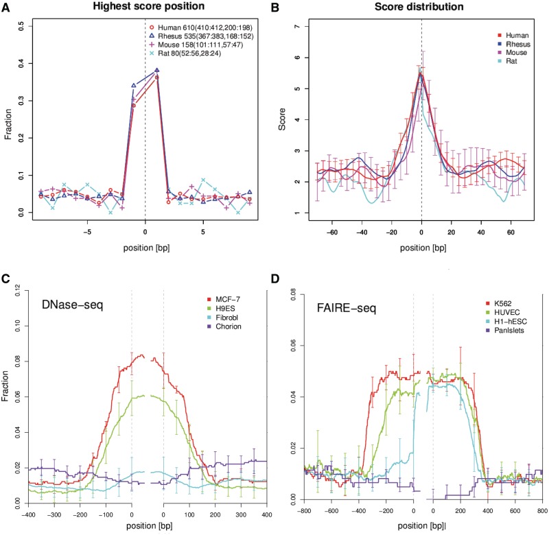
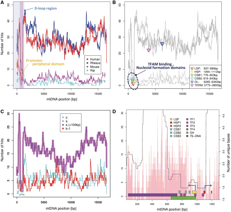
It is well known that remnants of partial or whole copies of mitochondrial DNA, known as Nuclear MiTochondrial sequences (NUMTs), are found in nuclear genomes. Since whole genome sequences have become available, many bioinformatics studies have identified putative NUMTs and from those attempted to infer the factors involved in NUMT creation. These studies conclude that NUMTs represent randomly chosen regions of the mitochondrial genome. There is less consensus regarding the nuclear insertion sites of NUMTs - previous studies have discussed the possible role of retrotransposons, but some recent ones have reported no correlation or even anti-correlation between NUMT sites and retrotransposons. These studies have generally defined NUMT sites using BLAST with default parameters. We analyze a redefined set of human NUMTs, computed with a carefully considered protocol. We discover that the inferred insertion points of NUMTs have a strong tendency to have high-predicted DNA curvature, occur in experimentally defined open chromatin regions and often occur immediately adjacent to A + T oligomers. We also show clear evidence that their flanking regions are indeed rich in retrotransposons. Finally we show that parts of the mitochondrial genome D-loop are under-represented as a source of NUMTs in primate evolution.


Figures

Figure 1.
Figure 1.
Features of NUMT insertion sites in the nuclear genome. (A) The proportion of NUMT flank bases covered by retrotransposons at each distance from 1 to 5000 bp is shown. (B) The relative ratio of 3–6 bp A+T-only oligomers in NUMT flanks is shown. The horizontal axis gives the position relative to the inferred NUMT boundary. The vertical axis gives the ratio of A+T-only oligomers centered at each flank position relative to the overall genome average. (C) Additional observations and discussion regarding NUMT insertion sites is depicted. We observe that the immediate flanking region of NUMTs tend to have A+T rich oligomers, high predicted DNA bendability and curvature and occur in open chromatin regions. From the enrichment of retrotransposons seen in (A) and other circumstantial evidence we speculate that L1 endonuclease nicking may have mediated many NUMT insertion events in primates.
Figure 2.
Figure 2.
Predicted DNA curvature in NUMT flanks. (A) The horizontal axis gives the distance from the inferred NUMT insertion site. The vertical axis gives the fraction of human NUMTs which attain a local maximum (within the 20 bp window shown) in predicted DNA curvature. (B) The score distribution of DNA curvature in concatenated NUMT flanks is shown. The vertical bars represent standard error. Clear peaks of curvature are observed at inferred NUMT insertion sites. Distributions of experimentally identified open chromatin regions with DNase-seq (C) and FAIRE-seq (D) in NUMT flanks. (C,D): The horizontal axis shows the distance from the inferred NUMT insertion sites. The vertical axis shows the fraction of human NUMTs which have open chromatin at each position.
Figure 3.
Figure 3.
The mitochondrial D-loop region tends not to form NUMTs. (AD): Histograms of NUMT frequencies. The horizontal axis indicates position in the mitochondrial genome and the vertical axis the number of NUMTs whose inferred source mtDNA overlaps at each position. Note that human mtDNA is circular, so the right edge of plots A–C are conceptually joined to the left edge. A the distribution in several species. The vertically shaded region denotes the D-loop region, with the portion under-represented in NUMTs highlighted in a darker shade. (B) Superimposes the binding positions of several mtDNA binding proteins. (C) The distribution of human NUMTs inserted in different ages. The region under-represented in NUMTs is indicated with a bar. (D) A close-up of the NUMT depleted D-loop region (LSP, light strand promoter, HSP, heavy strand promoter, CSB, conserved sequence block, TF, TFAM binding region, OH, heavy strand replication origin and 7S DNA, displacement loop). The arrows represent direction of transcription from LSP and HSP1. The pink histogram-like bars show a rough estimate of local NUMT detection hardness due to sequence divergence—the number of unique characters (base or gap) in a multiple alignment of the mtDNA of each organism used in this study, averaged over a window of four positions.

References

    1. Sagan L. On the origin of mitosing cells. J. Theol. Biol. 1967;14:255–274. Author later changed her name to Lynn Margulis. - PubMed
    1. du Buy H, Riley F. Hybridization between the nuclear and kinetoplast DNA's of Leishmania enriettii and between nuclear and mitochondrial DNA's of mouse liver. Proc. Natl Acad. Sci. USA. 1967;57:790–797. - PMC - PubMed
    1. Gherman A, Chen PE, Teslovich TM, Stankiewicz P, Withers M, Kashuk CS, Chakravarti A, Lupski JR, Cutler DJ, Katsanis N. Population bottlenecks as a potential major shaping force of human genome architecture. PLoS Genet. 2007;3:e119. - PMC - PubMed
    1. Hazkani-Covo E, Zeller RM, Martin W. Molecular Poltergeists: Mitochondrial DNA Copies (numts) in Sequenced Nuclear Genomes. PLoS Genet. 2010;6:e1000834. - PMC - PubMed
    1. Ricchetti M, Fairhead C, Dujon B. Mitochondrial DNA repairs double-strand breaks in yeast chromosomes. Nature. 1999;402:96–100. - PubMed

Publication types