. 2012 Aug 24;287(35):29801-14.
doi: 10.1074/jbc.M112.378281. Epub 2012 Jul 2.

Protein arginine methyltransferase 7 regulates cellular response to DNA damage by methylating promoter histones H2A and H4 of the polymerase δ catalytic subunit gene, POLD1

Affiliations

Protein arginine methyltransferase 7 regulates cellular response to DNA damage by methylating promoter histones H2A and H4 of the polymerase δ catalytic subunit gene, POLD1

Vrajesh Karkhanis et al. J Biol Chem. .

Abstract

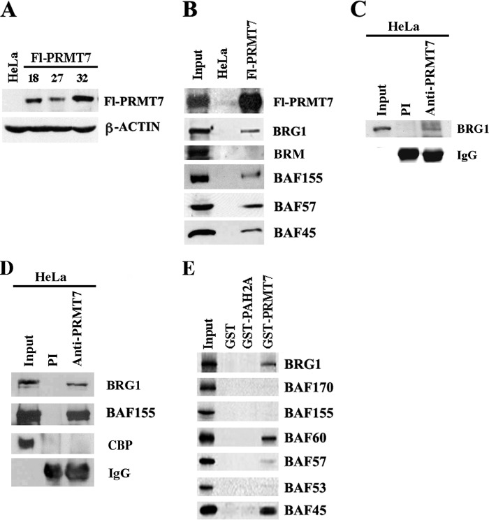
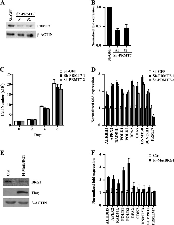
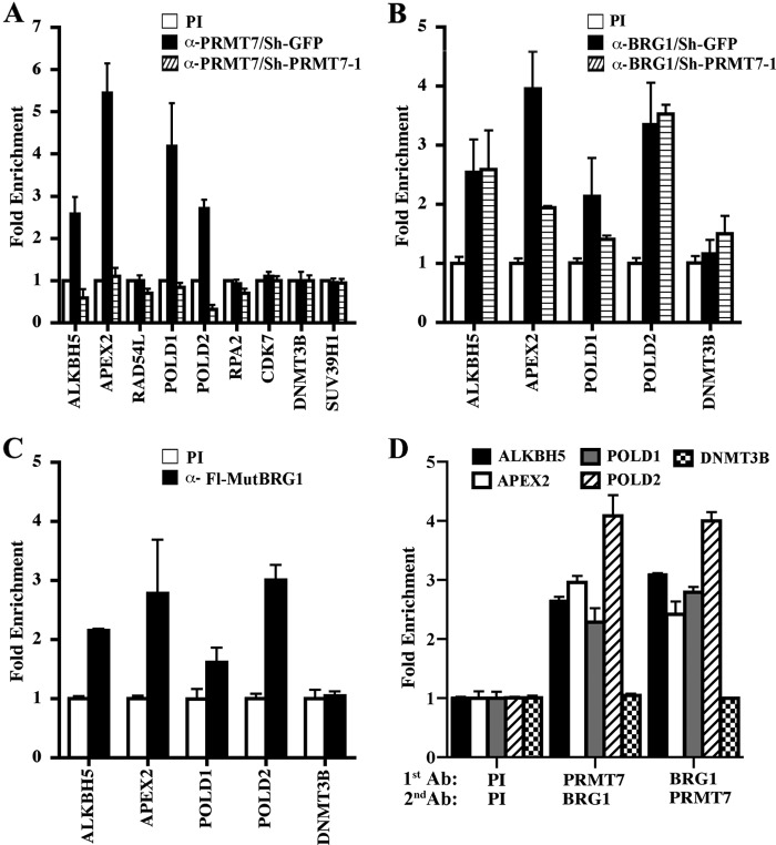
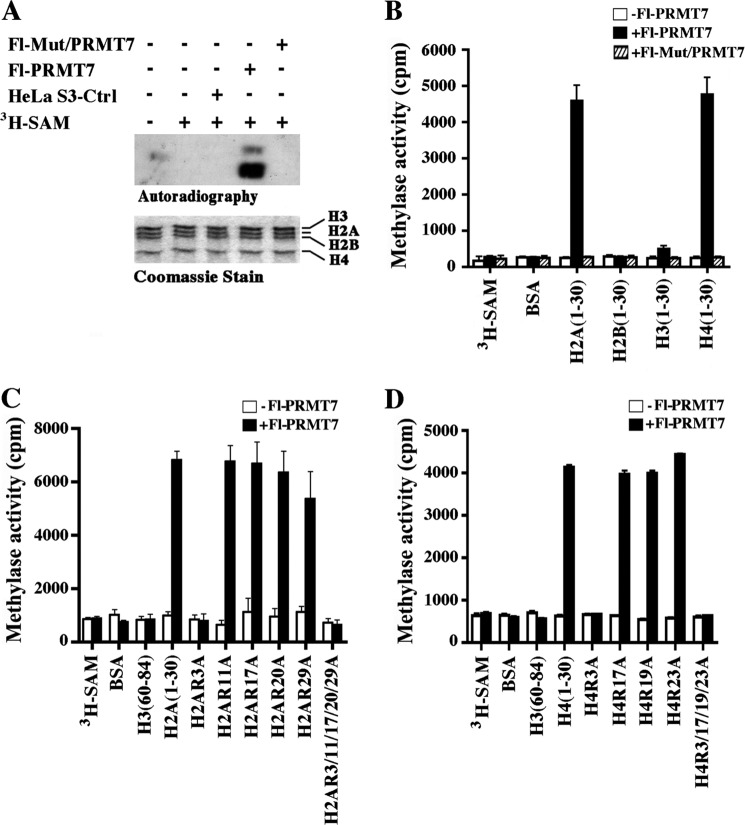
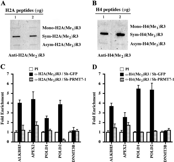
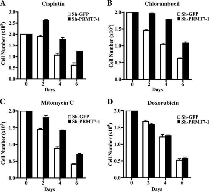
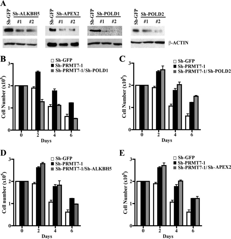
Covalent modification of histones by protein arginine methyltransferases (PRMTs) impacts genome organization and gene expression. In this report, we show that PRMT7 interacts with the BRG1-based hSWI/SNF chromatin remodeling complex and specifically methylates histone H2A Arg-3 (H2AR3) and histone H4 Arg-3 (H4R3). To elucidate the biological function of PRMT7, we knocked down its expression in NIH 3T3 cells and analyzed global gene expression. Our findings show that PRMT7 negatively regulates expression of genes involved in DNA repair, including ALKBH5, APEX2, POLD1, and POLD2. Chromatin immunoprecipitation (ChIP) revealed that PRMT7 and dimethylated H2AR3 and H4R3 are enriched at target DNA repair genes in parental cells, whereas PRMT7 knockdown caused a significant decrease in PRMT7 recruitment and H2AR3/H4R3 methylation. Decreased PRMT7 expression also resulted in derepression of target DNA repair genes and enhanced cell resistance to DNA-damaging agents. Furthermore, we show that BRG1 co-localizes with PRMT7 on target promoters and that expression of a catalytically inactive form of BRG1 results in derepression of PRMT7 target DNA repair genes. Remarkably, reducing expression of individual PRMT7 target DNA repair genes showed that only the catalytic subunit of DNA polymerase, POLD1, was able to resensitize PRMT7 knock-down cells to DNA-damaging agents. These results provide evidence for the important role played by PRMT7 in epigenetic regulation of DNA repair genes and cellular response to DNA damage.


Figures

FIGURE 1.
FIGURE 1.
PRMT7 can associate with BRG1-based hSWI/SNF. A, analysis of ectopically expressed FLAG-tagged PRMT7 (Fl-PRMT7) in stable HeLa S3 cell lines. Approximately 30 μg of nuclear extract from either HeLa S3 cells transfected with control vector (HeLa) or HeLa S3 clones 18, 27, and 32 stably transfected with pBabe/Fl-PRMT7 vector were analyzed by Western blotting using anti-FLAG antibody. Anti-β-ACTIN antibody was used as a control. B, components of the BRG1-based hSWI/SNF complex co-purify with Fl-PRMT7. An equal amount (15 μl) of peak fractions purified from control HeLa S3 or Fl-PRMT7 (clone 32) nuclear extract were analyzed by Western blotting using the indicated antibodies. Input represents 20 μg of Fl-PRMT7 crude nuclear extract. C, endogenous PRMT7 can interact with BRG1. Nuclear extract (250 mg) from HeLa S3 cells was immunoprecipitated using either preimmune (PI) (lane 2) or immune anti-PRMT7 antibody (lane 3), and bound proteins were analyzed by Western blot analysis using anti-BRG1 antibody. Input represents 20 mg of HeLa S3 nuclear extract. D, interaction between endogenous PRMT7 and BRG1-based hSWI/SNF is enhanced by the cross-linking agent dithiobis(succinimidyl propionate). Equal amounts of cross-linked HeLa S3 nuclear extract (250 mg) were incubated with PI or immune anti-PRMT7 antibody, and bound proteins were detected by immunoblotting using the indicated antibodies. E, specific BRG1-based hSWI/SNF subunits interact with PRMT7. In vitro-translated hSWI/SNF subunits were synthesized in the presence of [35S]methionine/cysteine using Promega TNT-coupled reticulocyte lysate, as described previously (14) and were incubated with either GST, GST-PAH2A, which includes the second paired amphipathic helix domain of mSIN3A, or GST-PRMT7. After extensive washing, the retained proteins were detected by autoradiography. Input represents 25% of the total amount of 35S-labeled protein used in each reaction. CBP, CREB-binding protein.
FIGURE 2.
FIGURE 2.
Effects of PRMT7 on cell growth and target gene expression. A, PRMT7 protein expression was measured by Western blot analysis using 30 μg of whole cell extract from control NIH 3T3/sh-GFP or PRMT7 knock-down cells, NIH 3T3/sh-PRMT7-1, and NIH 3T3/sh-PRMT7-2. Anti-β-ACTIN was used to show equal loading. B, PRMT7 mRNA levels were determined by real-time RT-PCR using 1 μg of total RNA from either control NIH 3T3/sh-GFP, NIH 3T3/sh-PRMT7-1, or NIH 3T3/sh-PRMT7-2. Real time RT-PCR was conducted three times in triplicate, and 18S rRNA was used as an internal control. C, growth rates of control and PRMT7 knock-down cell lines were measured by seeding 2 × 10⁵ cells in each plate, and the number of viable cells was determined by staining with trypan blue every 2 days for 6 days. The experiment was performed three times in triplicate, and the data points represent the average count from nine plates. D, PRMT7 regulates expression of genes involved in DNA repair. Real-time RT-PCR was performed on 1 μg of total RNA from control NIH 3T3/sh-GFP, NIH 3T3/sh-PRMT7-1, or NIH 3T3/sh-PRMT7-2 cells using primers specific for the indicated target genes. 18S rRNA levels were used as internal control. Each reaction was performed at least two times in triplicate. E, nuclear extract from control NIH 3T3 cells (Ctrl) or NIH 3T3 cells that expresses catalytically inactive FLAG-tagged BRG1 (Fl-MutBRG1) were analyzed by Western blotting using the indicated antibodies. F, PRMT7 target genes are transcriptionally derepressed in cells that express Fl-MutBRG1. mRNA levels of PRMT7 target genes were determined by real-time RT-PCR using 1 μg of total RNA from either Ctrl or Fl-MutBRG1 cells. Real-time RT-PCR was conducted three times in triplicate, and 18S rRNA was used as an internal control. Error bars show S.D.
FIGURE 3.
FIGURE 3.
PRMT7 is recruited to a select set of DNA repair genes. A, ChIP assays were carried out using cross-linked chromatin from control NIH 3T3/sh-GFP and NIH 3T3/sh-PRMT7-1 cells. Nucleoprotein complexes were immunoprecipitated with either PI or immune anti-PRMT7 antibodies, and bound DNA was analyzed by real-time PCR using gene-specific primers. -Fold enrichment was determined relative to the PI sample, and ChIP experiments were repeated three times in triplicate. B, BRG1 is recruited to the promoter region of PRMT7 target DNA repair genes. Cross-linked chromatin from control NIH 3T3/sh-GFP or NIH 3T3/sh-PRMT7-1 cells was immunoprecipitated with either PI or anti-BRG1 antibody, and the purified DNA was checked for enrichment of PRMT7 target promoters using gene-specific primers. C, catalytically inactive Fl-MutBRG1 is recruited to PRMT7 target genes. Chromatin from control NIH 3T3 or NIH 3T3 cells that express Fl-MutBRG1 was immunoprecipitated with either PI or anti-FLAG antibody, and DNA sequences of PRMT7 target promoters were detected by real-time PCR using gene-specific primers. D, PRMT7 and BRG1 co-localize on target DNA repair promoters. ChIP-re-ChIP assays were performed on cross-linked chromatin from NIH 3T3 cells using the indicated antibodies (1st Ab). Next, bound nucleoprotein complexes were released in the presence of 20 mm DTT, and subjected to a second immunoprecipitation (2nd Ab) using either PI, anti-PRMT7, or anti-BRG1. Real-time PCR was conducted as described above. Error bars show S.D.
FIGURE 4.
FIGURE 4.
Affinity-purified FLAG-tagged PRMT7 preferentially methylates histones H2A and H4. A, approximately 10 μl of affinity-purified peak fractions from either control HeLa S3, Fl-PRMT7, or Fl-Mut/PRMT7 cells were incubated with 2 μg of HeLa S3 core histones in the presence of [3H]AdoMet. Methylated histones were visualized by autoradiography, and Coomassie Blue staining was used to show equal amounts of core histones in each reaction. B, PRMT7 methylates H2A and H4 N-terminal tails. H2A, H2B, H3, and H4 peptides (2 μg) were incubated with or without affinity-purified Fl-PRMT7 and Fl-Mut/PRMT7 in the presence of [3H]AdoMet, and samples were spotted onto Whatman P-81 filter paper before methylation of histone peptides was quantified by liquid scintillation counting. To show specificity, methylation of BSA was also measured. C, wild type and mutant H2A peptides (2 μg) containing either single or multiple arginine to alanine substitutions at positions 3, 11, 17, 20, and 29 were incubated with or without affinity-purified Fl-PRMT7 in the presence of [3H]AdoMet, and methylation was measured as described in B. D, approximately 2 μg of either wild type or mutant H4 N-terminal peptides with single point mutations (R3A, R17A, R19A, or R23A) or mutations at all four positions were incubated with or without affinity-purified Fl-PRMT7 in the presence of [3H]AdoMet as described in C. Peptide methylation assays were performed in triplicate and repeated three times. Error bars show S.D.
FIGURE 5.
FIGURE 5.
PRMT7-induced epigenetic marks are enriched at target DNA repair genes. Anti-H2A(Me2)R3 (A) and anti-H4(Me2)R3 (B) antibodies are highly specific and do not cross-react with other methylation marks. Approximately 1 and 2 μg of either monomethylated, symmetrically methylated, or asymmetrically methylated H2AR3 and H4R3 peptides were slot-blotted on nitrocellulose, and anti-H2A(Me2)R3 and anti-H4(Me2)R3 antibodies were used to detect methylation marks. ChIP assays were conducted essentially as described in Fig. 3A using chromatin from control NIH 3T3/sh-GFP and NIH 3T3/sh-PRMT7-1 cells using either PI or immune anti-H2A(Me2)R3 (C) and anti-H4(Me2)R3 (D). DNA sequences subjected to ChIP were analyzed as described in Fig. 3A. Each ChIP experiment was repeated twice in triplicate. Error bars show S.D.
FIGURE 6.
FIGURE 6.
PRMT7 knock-down cells a more resistant to genotoxic stress. A–D, drug treatments were carried out as described under “Experimental Procedures” using an equal number (2 × 10⁵) of control NIH 3T3/sh-GFP and NIH 3T3/sh-PRMT7-1, and proliferation was monitored every 2 days for 6 days. The number of viable cells was determined by trypan blue staining. Each drug treatment was conducted twice in triplicate. Error bars show S.D.
FIGURE 7.
FIGURE 7.
Knockdown of the polymerase δ catalytic subunit POLD1 resensitizes PRMT7 knock-down cells to DNA damage. A, expression of PRMT7 target genes was knocked down using lentiviruses that express two distinct shRNAs (#1 and #2) against either ALKBH5, APEX2, POLD1, or POLD2. Protein expression of each PRMT7 target gene was evaluated by Western blot analysis using equal amounts of whole cell extracts (30 μg) from control NIH 3T3/sh-PRMT7-1 infected with lentivirus that expresses either sh-GFP or the indicated shRNAs against ALKBH5, APEX2, POLD1, and POLD2. PRMT7 target gene expression was determined using either anti-ALKBH5, anti-APEX2, anti-POLD1, or anti-POLD2 antibody. Anti-β-ACTIN was used a control. B–E, growth rates of control NIH 3T3/sh-GFP, NIH 3T3/sh-PRMT7-1, and NIH 3T3/sh-PRMT7-1 cells where expression of individual PRMT7 target DNA repair genes has been knocked down was measured by seeding 2 × 10⁵ cells in each plate and treating cells with cisplatin as described under “Experimental Procedures.” The number of viable cells was determined by trypan blue staining every 2 days for 6 days. Each experiment was conducted in triplicate. Error bars show S.D.

Similar articles

Cited by

References

    1. Bedford M. T., Clarke S. G. (2009) Protein arginine methylation in mammals. Who, what, and why. Mol. Cell 33, 1–13 - PMC - PubMed
    1. Cloos P. A., Christensen J., Agger K., Helin K. (2008) Erasing the methyl mark. Histone demethylases at the center of cellular differentiation and disease. Genes Dev. 22, 1115–1140 - PMC - PubMed
    1. Henikoff S. (2008) Nucleosome destabilization in the epigenetic regulation of gene expression. Nat. Rev. Genet. 9, 15–26 - PubMed
    1. Klose R. J., Zhang Y. (2007) Regulation of histone methylation by demethylimination and demethylation. Nat. Rev. Mol. Cell Biol. 8, 307–318 - PubMed
    1. Lee K. K., Workman J. L. (2007) Histone acetyltransferase complexes. One size doesn't fit all. Nat. Rev. Mol. Cell Biol. 8, 284–295 - PubMed

Publication types

MeSH terms

Substances

LinkOut - more resources