

STAT3 protein interacts with Class O Forkhead transcription factors in the cytoplasm and regulates nuclear/cytoplasmic localization of FoxO1 and FoxO3a proteins in CD4(+) T cells
- PMID: 22761423
- PMCID: PMC3436293
- DOI: 10.1074/jbc.M112.359661
STAT3 protein interacts with Class O Forkhead transcription factors in the cytoplasm and regulates nuclear/cytoplasmic localization of FoxO1 and FoxO3a proteins in CD4(+) T cells
Abstract
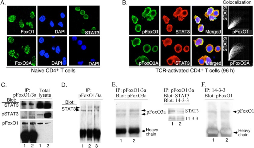
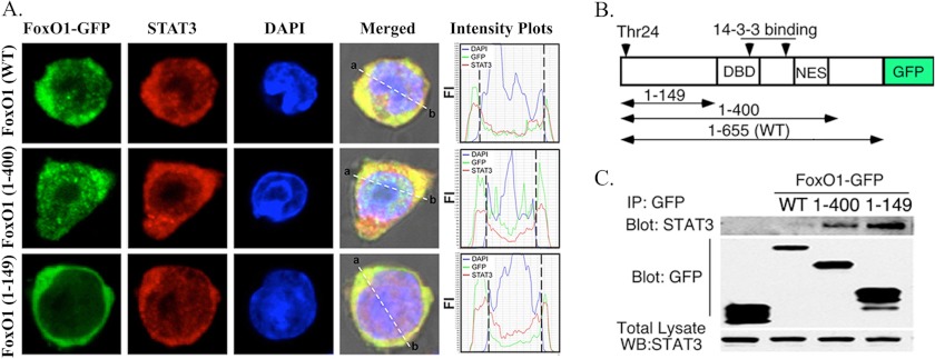
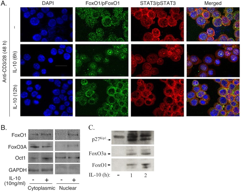
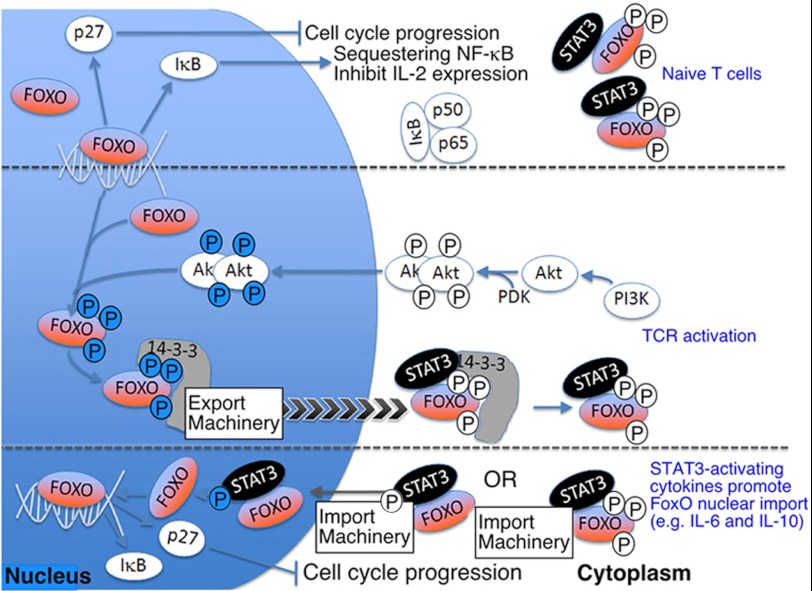
An important feature of the adaptive immune response is its remarkable capacity to regulate the duration of inflammatory responses, and effector T cells have been shown to limit excessive immune responses by producing anti-inflammatory cytokines such as IL-10 and IL-27. However, how anti-inflammatory cytokines mediate their suppressive activities is not well understood. In this study, we show that STAT3 contributes to mechanisms that control the duration of T cell proliferation by regulating the subcellular location of FoxO1 and FoxO3a, two Class O Forkhead transcription factors that mediate lymphocyte quiescence and inhibit T cell activation. We show that active FoxO1 and FoxO3a reside exclusively in the nucleus of naïve T cells whereas inactive pFoxO1 and pFoxO3a were most abundant in activated T cells and sequestered in their cytoplasm in association with unphosphorylated STAT3 (U-STAT3) and 14-3-3. We further show that FoxO1/FoxO3a rapidly relocalized into the nucleus in response to pSTAT3 activation by IL-6 or IL-10, and the accumulation of FoxO1/FoxO3a in their nuclei coincided with increased expression of p27(Kip1) and p21(WAF1). STAT3 inhibitors completely abrogated cytokine-induced translocation of FoxO1/FoxO3a into the nucleus. In naïve or resting STAT3-deficient T cells, expression of pFoxO1/pFoxO3a was predominantly in the cytoplasm and correlated with defects in p27(Kip1) and p21(WAF1) expression, suggesting requirement of STAT3 for importation or retention of FoxO in the nucleus and attenuation of lymphocyte proliferation. Taken together, these results suggest that U-STAT3 collaborates with 14-3-3 to sequester pFoxO1/pFoxO3a in cytoplasm and thus prolong T cell activation, whereas pSTAT3 activation by anti-inflammatory cytokines would curtail the duration of TCR activation and re-establish lymphocyte quiescence by inducing nuclear localization of FoxO1/FoxO3a and FoxO-mediated expression of growth-inhibitory proteins.
Figures


Similar articles
-
STAT3 protein promotes T-cell survival and inhibits interleukin-2 production through up-regulation of Class O Forkhead transcription factors.J Biol Chem. 2011 Sep 2;286(35):30888-30897. doi: 10.1074/jbc.M111.253500. Epub 2011 Jul 5. J Biol Chem. 2011. PMID: 21730069 Free PMC article.
-
The inhibition of activated hepatic stellate cells proliferation by arctigenin through G0/G1 phase cell cycle arrest: persistent p27(Kip1) induction by interfering with PI3K/Akt/FOXO3a signaling pathway.Eur J Pharmacol. 2015 Jan 15;747:71-87. doi: 10.1016/j.ejphar.2014.11.040. Epub 2014 Dec 10. Eur J Pharmacol. 2015. PMID: 25498792
-
Kinetics of nuclear-cytoplasmic translocation of Foxo1 and Foxo3A in adult skeletal muscle fibers.Am J Physiol Cell Physiol. 2012 Nov 1;303(9):C977-90. doi: 10.1152/ajpcell.00027.2012. Epub 2012 Aug 29. Am J Physiol Cell Physiol. 2012. PMID: 22932683 Free PMC article.
-
FoxO1 - the key for the pathogenesis and therapy of acne?J Dtsch Dermatol Ges. 2010 Feb;8(2):105-14. doi: 10.1111/j.1610-0387.2010.07344.x. J Dtsch Dermatol Ges. 2010. PMID: 20151947 Review. English, German.
-
Foxo3a: an integrator of immune dysfunction during HIV infection.Cytokine Growth Factor Rev. 2012 Aug-Oct;23(4-5):215-21. doi: 10.1016/j.cytogfr.2012.05.008. Epub 2012 Jun 27. Cytokine Growth Factor Rev. 2012. PMID: 22748238 Free PMC article. Review.
Cited by
-
[8] and [10]-Gingerol reduces urothelial damage in ifosfamide-induced hemorrhagic cystitis via JAK/STAT/FOXO signaling pathway via IL-10.Naunyn Schmiedebergs Arch Pharmacol. 2023 Aug;396(8):1773-1786. doi: 10.1007/s00210-023-02436-2. Epub 2023 Feb 27. Naunyn Schmiedebergs Arch Pharmacol. 2023. PMID: 36843129
-
Blueberry extract attenuates norepinephrine-induced oxidative stress and apoptosis in H9c2 cardiac cells.Mol Cell Biochem. 2022 Mar;477(3):663-672. doi: 10.1007/s11010-021-04313-z. Epub 2022 Jan 6. Mol Cell Biochem. 2022. PMID: 34988854
-
STAT3-Specific Single Domain Nanobody Inhibits Expansion of Pathogenic Th17 Responses and Suppresses Uveitis in Mice.Front Immunol. 2021 Sep 15;12:724609. doi: 10.3389/fimmu.2021.724609. eCollection 2021. Front Immunol. 2021. PMID: 34603297 Free PMC article.
-
SMAD3/Stat3 Signaling Mediates β-Cell Epithelial-Mesenchymal Transition in Chronic Pancreatitis-Related Diabetes.Diabetes. 2017 Oct;66(10):2646-2658. doi: 10.2337/db17-0537. Epub 2017 Aug 3. Diabetes. 2017. PMID: 28775125 Free PMC article.
-
STAT3 and its targeting inhibitors in osteosarcoma.Cell Prolif. 2021 Feb;54(2):e12974. doi: 10.1111/cpr.12974. Epub 2020 Dec 31. Cell Prolif. 2021. PMID: 33382511 Free PMC article. Review.
References
-
- Liu J. O. (2005) The yins of T cell activation. Sci. STKE 2005, re1. - PubMed
-
- Coffer P. J., Burgering B. M. (2004) Forkhead-box transcription factors and their role in the immune system. Nat. Rev. Immunol. 4, 889–899 - PubMed
-
- Brunet A., Bonni A., Zigmond M. J., Lin M. Z., Juo P., Hu L. S., Anderson M. J., Arden K. C., Blenis J., Greenberg M. E. (1999) Akt promotes cell survival by phosphorylating and inhibiting a Forkhead transcription factor. Cell 96, 857–868 - PubMed
-
- Lin L., Hron J. D., Peng S. L. (2004) Regulation of NF-κB, Th activation, and autoinflammation by the Forkhead transcription factor FoxO3a. Immunity 21, 203–213 - PubMed
-
- Stahl M., Dijkers P. F., Kops G. J., Lens S. M., Coffer P. J., Burgering B. M., Medema R. H. (2002) The Forkhead transcription factor FoxO regulates transcription of p27Kip1 and Bim in response to IL-2. J. Immunol. 168, 5024–5031 - PubMed
Publication types
MeSH terms
Substances
Grants and funding
LinkOut - more resources
Full Text Sources
Other Literature Sources
Molecular Biology Databases
Research Materials
Miscellaneous
