. 2012 Sep 14;287(38):32054-68.
doi: 10.1074/jbc.M112.371476. Epub 2012 Jul 2.

Polycyclic aromatic hydrocarbons (PAHs) mediate transcriptional activation of the ATP binding cassette transporter ABCB6 gene via the aryl hydrocarbon receptor (AhR)

Affiliations

Polycyclic aromatic hydrocarbons (PAHs) mediate transcriptional activation of the ATP binding cassette transporter ABCB6 gene via the aryl hydrocarbon receptor (AhR)

Hemantkumar Chavan et al. J Biol Chem. .

Abstract

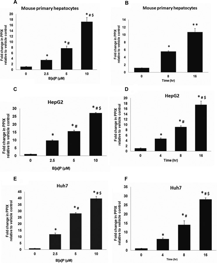
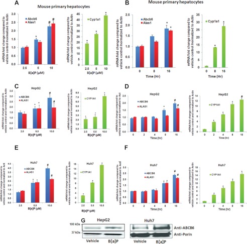
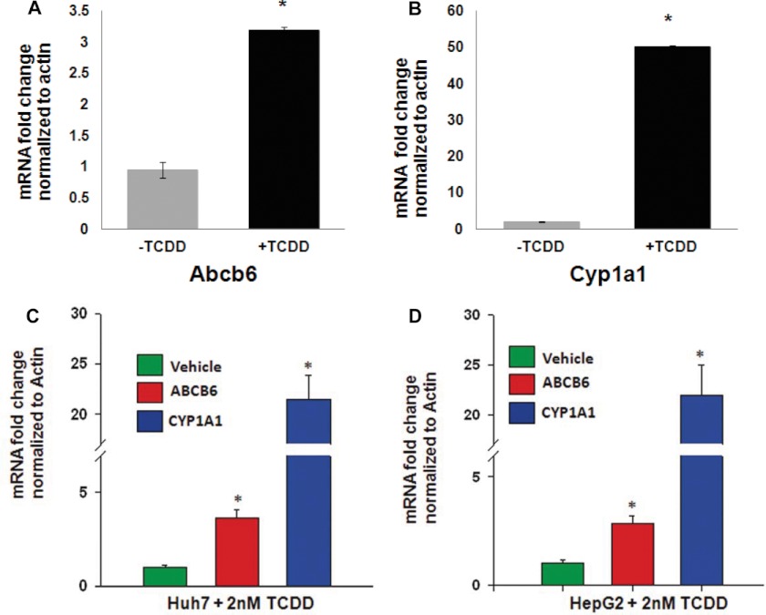
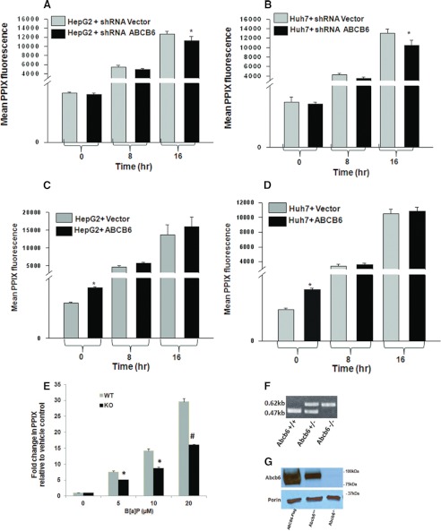
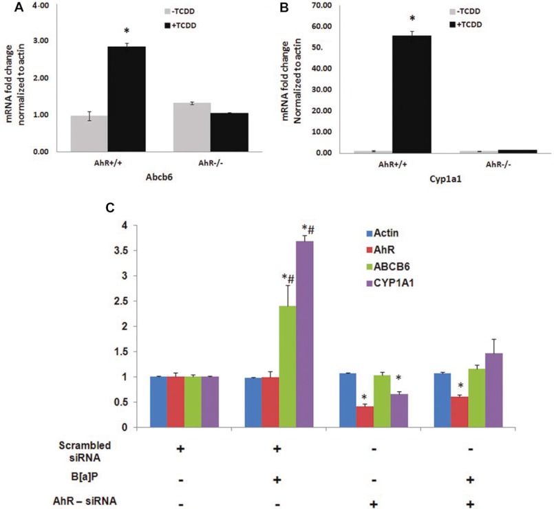
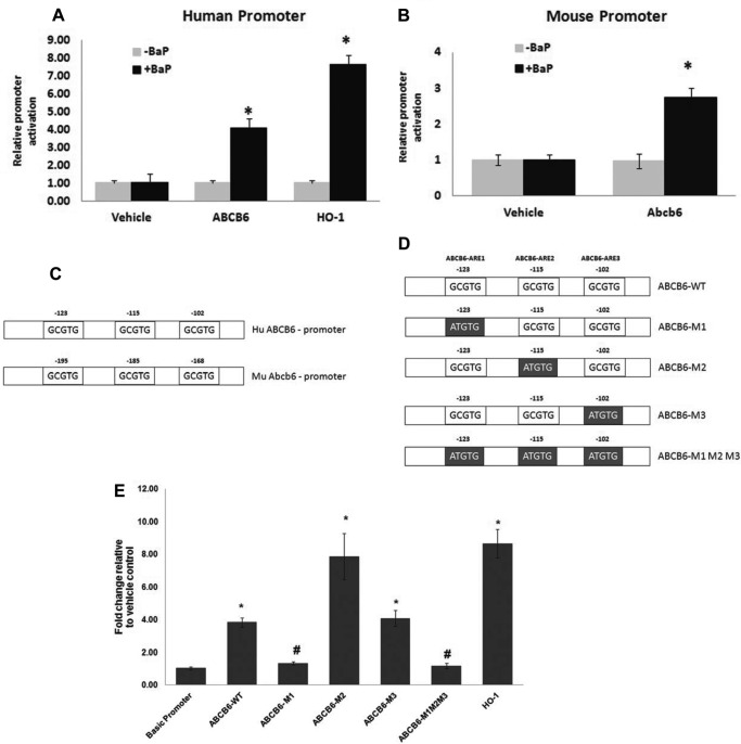
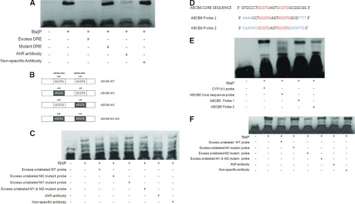
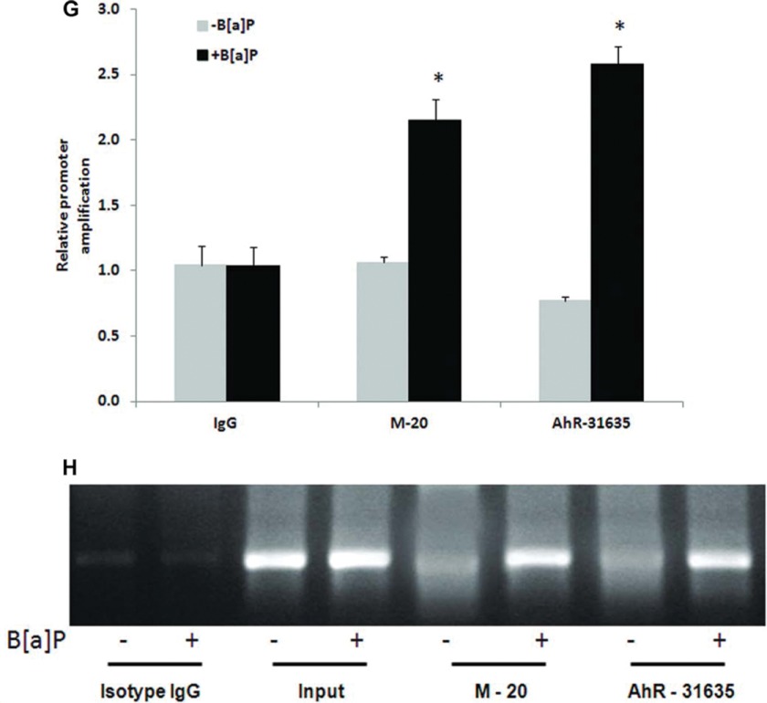
Liver is endowed with a mechanism to induce hepatic cytochromes P450 (CYP450s) in response to therapeutic drugs and environmental contaminants, leading to increased detoxification and elimination of the xenobiotics. Each CYP450 is composed of an apoprotein moiety and a heme prosthetic group, which is required for CYP450 activity. Thus, under conditions of CYP450 induction, there is a coordinate increase in heme biosynthesis to compensate for the increased expression of CYP450s. ABCB6, a mitochondrial ATP binding cassette transporter, which regulates coproporphyrinogen transport from the cytoplasm into the mitochondria to complete heme biosynthesis, represents a previously unrecognized rate-limiting step in heme biosynthesis. However, it is not known if exposure to drugs and environmental contaminants induces ABCB6 expression, to assure an adequate and apparently coordinated supply of heme for the generation of functional cytochrome holoprotein. In the present study, we demonstrate that polycyclic aromatic hydrocarbons (PAHs), the widely distributed environmental toxicants shown to induce porphyrin accumulation causing hepatic porphyria, up-regulate ABCB6 expression in both mice and humans. Using siRNA technology and Abcb6 knock-out mice, we demonstrate that PAH-mediated increase in hepatic porphyrins is compromised in the absence of ABCB6. Moreover, in vivo studies in aryl hydrocarbon receptor (AhR) knock-out mice demonstrate that PAH induction of ABCB6 is mediated by AhR. Promoter activation studies combined with electrophoretic mobility shift assay and chromatin immunoprecipitation assay demonstrate direct interactions between the AhR binding sites in the ABCB6 promoter and the AhR receptor, implicating drug activation mechanisms for ABCB6 similar to those found in inducible cytochrome P450s. These studies are the first to describe direct transcriptional activation of both mouse and human ABCB6 by xenobiotics.


Figures

FIGURE 1.
FIGURE 1.
Polyaromatic hydrocarbon benzo[a]pyrene increases hepatic protoporphyrin IX levels. Exposure to B[a]P increases protoporphyrin IX levels in both mouse primary hepatocytes (A and B) and human hepatomas (C–F), in a dose-dependent (A, C, and E) and time-dependent (B, D, and F) manner. Values represent mean ± S.D. (error bars) (n = 4). Results shown are representative of three independent experiment with n = 4/experiment. *, significantly different from vehicle control (p < 0.01); #, significantly different from cells treated with 2.5 μm B[a]P in dose-dependent studies and significantly different from 4-h treatment (hepatomas) in time-dependent studies (p < 0.01). $, significantly different from cells treated with 5 μm B[a]P in dose-dependent studies and significantly different from 8-h treatment (hepatomas) in time-dependent studies (p < 0.01). **, significantly different from cells exposed to B[a]P for 8 h (mouse primary hepatocytes) (p < 0.01).
FIGURE 2.
FIGURE 2.
Benzo[a]pyrene induces ABCB6 and ALAS1 expression in a dose- and time-dependent manner. Exposure to B[a]P induces Abcb6 and Alas1 expression in mouse primary hepatocytes (left panels; A and B) and human hepatomas (left panels; C–F) in a dose-dependent (left panels; A, C, and E) and time-dependent manner (left panels; B, D, and F). Right panels (A–F), B[a]P-mediated induction of CYP1A1. CYP1A1, a gene known to be induced by B[a]P in liver, is used as a positive control in these experiments. G, ABCB6 protein expression in response to B[a]P treatment in human hepatomas. ABCB6 expression was measured in isolated mitochondria using an ABCB6-specific antibody. Values represent mean ± S.D. (error bars) (n = 4). Results shown are representative of three independent experiments with n = 4/experiment. *, significantly different from cells exposed to 2.5 μm B[a]P in dose-dependent studies and significantly different from cells exposed to B[a]P for 4 h (hepatomas) in time-dependent studies (p < 0.01). #, significantly different from cells treated with 5 μm B[a]P in dose-dependent studies and significantly different from 8-h treatment (hepatomas) in time-dependent studies (p < 0.01).
FIGURE 3.
FIGURE 3.
TCDD induces ABCB6 expression in both mice and humans. Exposure to TCDD induces Abcb6 expression in mouse (A) and human hepatomas (C and D). B, TCDD-mediated induction of Cyp1a1, a gene known to be induced by TCDD in the liver, is used as a positive control in these experiments. Values represent mean ± S.D. (error bars); n = 6 for mice, and n = 4 for cell culture. Results shown are representative of three independent experiments. *, significantly different for vehicle-treated mice in animals and human hepatomas (p < 0.01).
FIGURE 4.
FIGURE 4.
Loss of ABCB6 expression compromises B[a]P-induced hepatic porphyrin levels. Endogenous knockdown of ABCB6 expression results in decreased protoporphyrin IX levels in both HepG2 (A) and Huh7 (B) cells exposed to B[a]P. In contrast, exposure to B[a]P does not affect protoporphyrin IX levels in HepG2 (C) and Huh7 (D) cells overexpressing ABCB6. E, loss of Abcb6 expression results in decreased protoporphyrin IX levels in mouse primary hepatocytes isolated from Abcb6−/− mice exposed to B[a]P. Genotyping (F) and Western blot (G) demonstrate loss of Abcb6 expression in Abcb6−/− mice. Values represent mean ± S.D. (error bars) (n = 3). Results are representative of three independent experiments with n = 3 per experiment. *, significantly different from shRNA vector control cells exposed to B[a]P for 16 h in (A and B) and significantly different from vector control cells (C and D). p < 0.05. E, *, significantly different from B[a]P-treated wild-type mouse primary hepatocytes. p < 0.01. #, significantly different from B[a]P-treated wild-type mouse primary hepatocytes. p < 0.001.
FIGURE 5.
FIGURE 5.
A functional AhR pathway is required for B[a]P-mediated up-regulation of ABCB6. Mouse (A) and HepG2 (C) cells lacking AhR are unable to up-regulate Abcb6 expression, whereas mouse (A) and HepG2 (C) cells with functional AhR up-regulate Abcb6 expression in response to B[a]P. B, B[a]P-mediated induction of Cyp1a1, a gene known to be induced by B[a]P, is used as a positive control in these experiments. Values represent mean ± S.D. (error bars); n = 4 for mice, and n = 3 for HepG2. Results representative of three independent experiments. *, significantly different from scrambled siRNA transfected cells; p < 0.01 in hepatomas and AhR+/+ vehicle-treated mice. #, significantly different from AhR siRNA-transfected cells treated with B[a]P. p < 0.01.
FIGURE 6.
FIGURE 6.
Active AhR response element in human and mouse 5′-flanking region. Shown is activity of human (A) and mouse (B) 5′-flanking sequence in the Abcb6 gene in response to B[a]P. C, schematic representation of the 5′-flanking region of human and mouse Abcb6 genes. Core AhREs are shown as boxes with respective locations of the 5′ end base from the reported transcription start sites. D, schematic representation of the human ABCB6 promoter, used in the transactivation studies, showing the introduction of mutations in the AhRE. Gray shaded boxes show the AhR sequence motif that was mutated by site-directed mutagenesis on the human ABCB6 promoter. E, mutation of either the distal AhRE or all of the three AhREs does not activate the promoter-luciferase reporter in response to B[a]P. Values represent mean ± S.D. (error bars) (n = 3). Results representative of six independent experiments. *, significantly different from empty vector-transfected cells treated with B[a]P and ABCB6 promoter-transfected cells treated with vehicle. p < 0.01. #, significantly different from ABCB6-WT, ABCB6-M1, and ABCB6-M2 promoter-transfected cells treated with B[a]P. p < 0.01.
FIGURE 7.
FIGURE 7.
AhR is recruited to the distal AhR response element in the human ABCB6 promoter. A, electrophoretic mobility shift assay of AhR complex binding to the AhRE element in the CYP1A1 promoter. B, schematic representation of the human ABCB6 promoter used in EMSA studies. C, electrophoretic mobility shift assay of AhR complex binding to the AhRE element in the human ABCB6 promoter. D, schematic representation of the changes introduced in the AhRE flanking sequence. Letters highlighted in blue show the altered bases introduced in the AhRE flanking sequence, and letters in red show the core AhRE. E and F, electrophoretic mobility shift assay of AhR complex binding to the human ABCB6-WT promoter and to the human ABCB6 promoter carrying changes to the AhRE flanking sequence. In all EMSA assays (A, C, E, and F), biotinylated DNA probes containing AhRE were incubated with nuclear extracts of HepG2 cells treated with either vehicle or 5 μm B[a]P in the presence or absence of excess unlabeled probe or the mutant AhREs. Polyclonal antibody against AhR was used to show antibody-induced reduction in the intensity of the AhR-protein complex band. Incubation with equal amounts of a nonspecific polyclonal antibody did not reduce the band intensity. Results are representative of four independent experiments. G and H, chromatin immunoprecipitation analysis shows recruitment of AhR to the AhRE in the ABCB6 promoter. Histograms (G) represent real-time PCR values of promoter amplification, whereas H shows conventional PCR analysis (28 cycles) for AhREs of ABCB6 to confirm the specificity of PCR amplification and quantitation in SYBR green real-time PCR. Immunoprecipitation was carried out with two anti-AhR antibodies (M-20 and AhR-31635) or isotype control IgG. Values represent mean ± S.D. (error bars) of three independent experiments. *, significantly different from B[a]P-treated isotype IgG control values. p < 0.01.
FIGURE 7.
FIGURE 7.
AhR is recruited to the distal AhR response element in the human ABCB6 promoter. A, electrophoretic mobility shift assay of AhR complex binding to the AhRE element in the CYP1A1 promoter. B, schematic representation of the human ABCB6 promoter used in EMSA studies. C, electrophoretic mobility shift assay of AhR complex binding to the AhRE element in the human ABCB6 promoter. D, schematic representation of the changes introduced in the AhRE flanking sequence. Letters highlighted in blue show the altered bases introduced in the AhRE flanking sequence, and letters in red show the core AhRE. E and F, electrophoretic mobility shift assay of AhR complex binding to the human ABCB6-WT promoter and to the human ABCB6 promoter carrying changes to the AhRE flanking sequence. In all EMSA assays (A, C, E, and F), biotinylated DNA probes containing AhRE were incubated with nuclear extracts of HepG2 cells treated with either vehicle or 5 μm B[a]P in the presence or absence of excess unlabeled probe or the mutant AhREs. Polyclonal antibody against AhR was used to show antibody-induced reduction in the intensity of the AhR-protein complex band. Incubation with equal amounts of a nonspecific polyclonal antibody did not reduce the band intensity. Results are representative of four independent experiments. G and H, chromatin immunoprecipitation analysis shows recruitment of AhR to the AhRE in the ABCB6 promoter. Histograms (G) represent real-time PCR values of promoter amplification, whereas H shows conventional PCR analysis (28 cycles) for AhREs of ABCB6 to confirm the specificity of PCR amplification and quantitation in SYBR green real-time PCR. Immunoprecipitation was carried out with two anti-AhR antibodies (M-20 and AhR-31635) or isotype control IgG. Values represent mean ± S.D. (error bars) of three independent experiments. *, significantly different from B[a]P-treated isotype IgG control values. p < 0.01.

Similar articles

Cited by

References

    1. Ponka P. (1999) Cell biology of heme. Am. J. Med. Sci. 318, 241–256 - PubMed
    1. Padmanaban G., Venkateswar V., Rangarajan P. N. (1989) Heme as a multifunctional regulator. Trends Biochem. Sci. 14, 492–496 - PubMed
    1. Sassa S., Nagai T. (1996) The role of heme in gene expression. Int. J. Hematol. 63, 167–178 - PubMed
    1. Aft R. L., Mueller G. C. (1984) Hemin-mediated oxidative degradation of proteins. J. Biol. Chem. 259, 301–305 - PubMed
    1. Maines M. D., Kappas A. (1975) The degradative effects of porphyrins and heme compounds on components of the microsomal mixed function oxidase system. J. Biol. Chem. 250, 2363–2369 - PubMed

Publication types

MeSH terms

LinkOut - more resources