Clinical Trial
. 2012 Aug 24;287(35):29261-72.
doi: 10.1074/jbc.M112.366518. Epub 2012 Jul 2.

Keratinization-associated miR-7 and miR-21 regulate tumor suppressor reversion-inducing cysteine-rich protein with kazal motifs (RECK) in oral cancer

Affiliations
Clinical Trial

Keratinization-associated miR-7 and miR-21 regulate tumor suppressor reversion-inducing cysteine-rich protein with kazal motifs (RECK) in oral cancer

Hyun Min Jung et al. J Biol Chem. .

Abstract

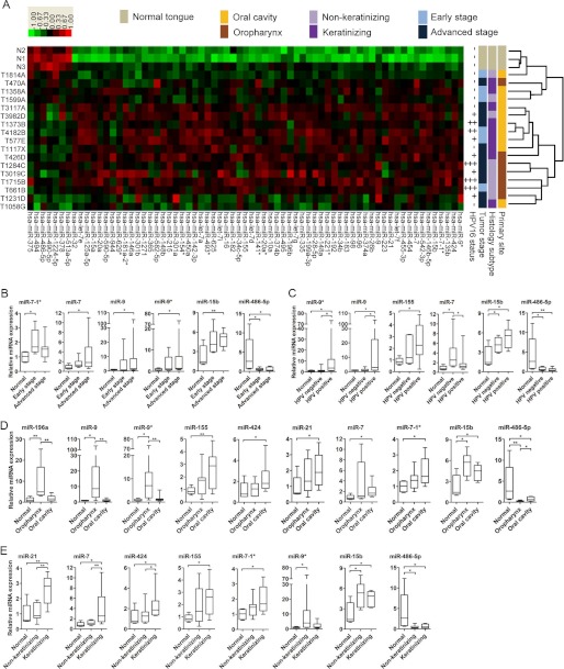
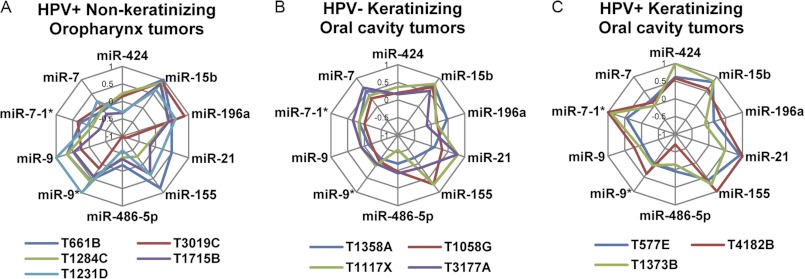
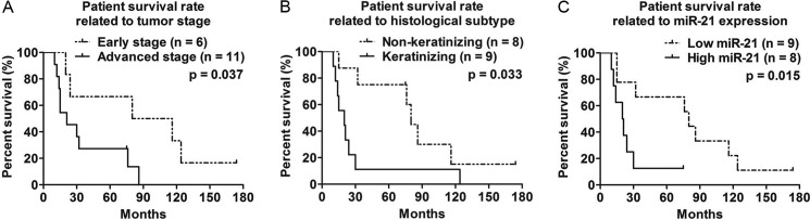
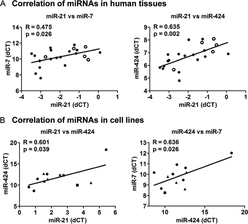
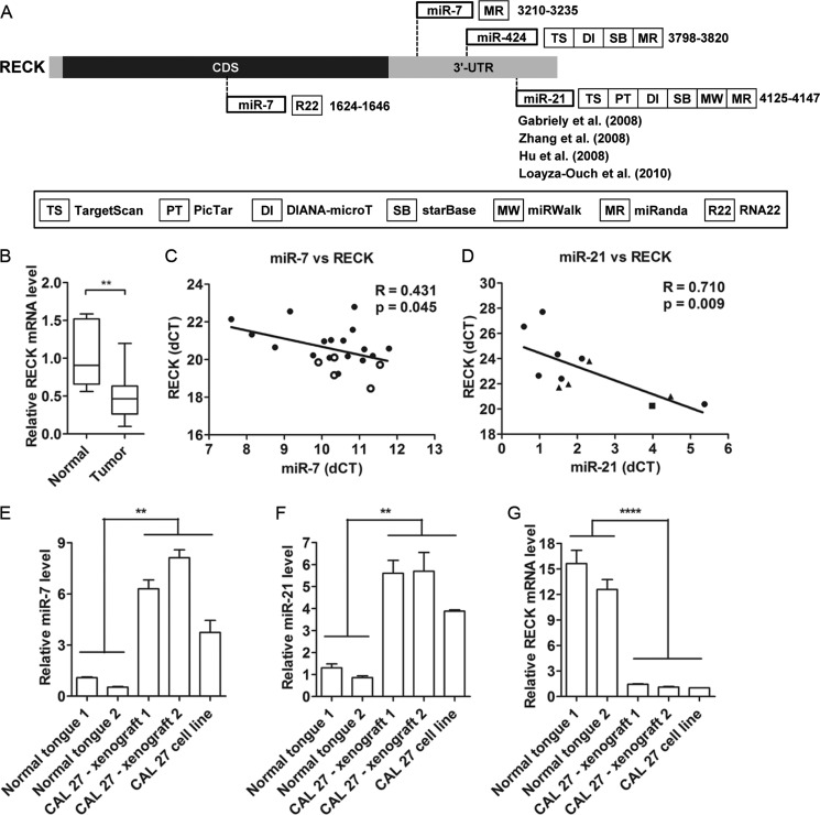
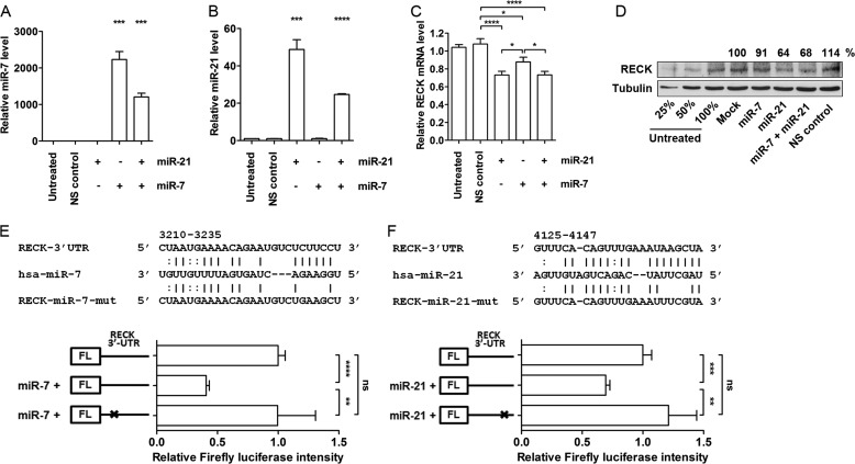
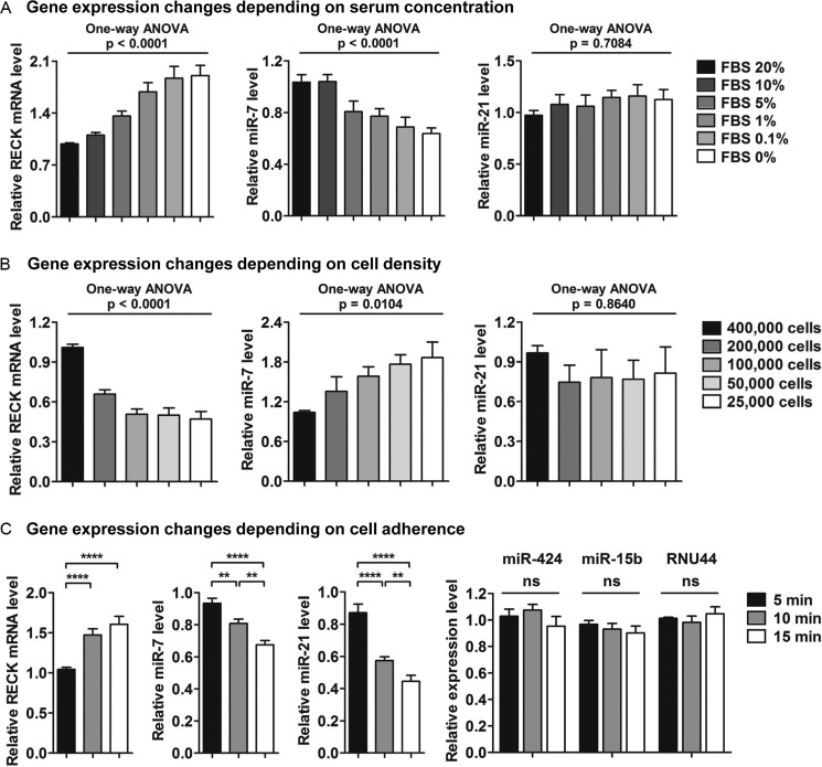
MicroRNAs (miRNAs) are small non-coding RNAs that posttranscriptionally regulate gene expression during many biological processes. Recently, the aberrant expressions of miRNAs have become a major focus in cancer research. The purpose of this study was to identify deregulated miRNAs in oral cancer and further focus on specific miRNAs that were related to patient survival. Here, we report that miRNA expression profiling provided more precise information when oral squamous cell carcinomas were subcategorized on the basis of clinicopathological parameters (tumor primary site, histological subtype, tumor stage, and HPV16 status). An innovative radar chart analysis method was developed to depict subcategories of cancers taking into consideration the expression patterns of multiple miRNAs combined with the clinicopathological parameters. Keratinization of tumors and the high expression of miR-21 were the major factors related to the poor prognosis of patients. Interestingly, a majority of the keratinized tumors expressed high levels of miR-21. Further investigations demonstrated the regulation of the tumor suppressor gene reversion-inducing cysteine-rich protein with kazal motifs (RECK) by two keratinization-associated miRNAs, miR-7 and miR-21. Transfection of miR-7 and miR-21-mimics reduced the expression of RECK through direct miRNA-mediated regulation, and these miRNAs were inversely correlated with RECK in CAL 27 orthotopic xenograft tumors. Furthermore, a similar inverse correlation was demonstrated in CAL 27 cells treated in vitro by different external stimuli such as trypsinization, cell density, and serum concentration. Taken together, our data show that keratinization is associated with poor prognosis of oral cancer patients and keratinization-associated miRNAs mediate deregulation of RECK which may contribute to the aggressiveness of tumors.


Figures

FIGURE 1.
FIGURE 1.
Differential miRNA expression in OSCC subtypes. A, 17 OSCC tumors form clusters on the basis of miRNA expression patterns and clinicopathological parameters. Heat maps were created by supervised hierarchical clustering analysis of 69 miRNAs (p < 0.05) with Log2 difference of > 1.5 between 17 OSCCs and three normal tongue samples. miRNA expression (columns) is shown for individual human tissue samples (rows). Four different clinicopathological classifications were applied to characterize the tumors: primary site (yellow/brown), histological subtype (light purple/dark purple), tumor stage (light blue/dark blue), and HPV16 status (−, +, ++, or +++). Gray represents normal tongue samples (N1-N3). Red and green colors in the heat map indicate overexpression and underexpression, respectively. Differential expressions of 10 miRNAs were verified in subtypes of OSCCs using Taqman qRT-PCR. Tumor samples were separated into subtypes on the basis of clinicopathological parameters: early (n = 6) versus advanced stage (n = 11) (B); HPV-negative (n = 7) versus HPV-positive (n = 10) (C); oropharynx (n = 7) versus oral cavity (n = 10) tumors (D); non-keratinizing (n = 8) versus keratinizing (n = 9) tumors (E). In addition to the three normal tissues used for the microarray analyses, two normal tongue tissues were added in the qRT-PCR analyses to enhance statistical computation. All qRT-PCR results are expressed as mean ± S.E. from at least three independent experiments. The results were analyzed using Mann-Whitney U test. *, p < 0.05, **, p < 0.01.
FIGURE 2.
FIGURE 2.
Combined clinicopathological parameters and miRNA expression signatures for OSCC subtypes. Similar contours of miRNA expression patterns are formed distinguishing HPV-positive non-keratinizing oropharynx tumors (A), HPV-negative keratinizing oral cavity tumors (B), and HPV-positive keratinizing oral cavity tumors (C).
FIGURE 3.
FIGURE 3.
Advanced tumor stage, keratinization state, and high expression of miR-21 as indicators of poor prognosis for oral cancer patients. Kaplan-Meier curves with Log-rank test were applied to identify the factors related to prognosis. A, advanced stage (p = 0.037). B, keratinization (p = 0.033). C, high expression of miR-21 (p = 0.015) were associated with poor survival rate.
FIGURE 4.
FIGURE 4.
Coordinated expression of keratinization-associated miRNAs in human tissues and cell lines. A, linear regression analyses of miRNAs in human oral tissues. Positive correlation was observed between miR-21 and miR-7 (r = 0.475, p = 0.026) and miR-21 and miR-424 (r = 0.635, p = 0.002). ● and ○ indicate 17 OSCCs and 5 normal tongue samples, respectively. B, linear regression analyses of miRNAs in human cancer cell lines. Positive correlation was observed between miR-21 and miR-424 (r = 0.601, p = 0.039) and miR-424 and miR-7 (r = 0.636, p = 0.026). ●, ▴, and ■ indicate head and neck cancer, cervical cancer, and HEK293 cells, respectively.
FIGURE 5.
FIGURE 5.
RECK is a common target of keratinization-associated miRNAs. A, in silico analysis identified a candidate target for miR-7, miR-21, and miR-424. Seven different miRNA target prediction programs were used in this analysis. B, RECK mRNA expression was measured in human oral tissues using Taqman qPCR. C, expression of miR-7 was inversely correlated with RECK in human oral tissues (r = 0.431, p = 0.045). ● and ○ indicate 17 OSCCs and five normal samples, respectively. D, expression of miR-21 was inversely correlated with RECK in human cancer cell lines. ●, ▴, and ■ indicate head and neck cancer cells, cervical cancer cells, and HEK293 cells, respectively. miR-7 (E) and miR-21 (F) expression levels were elevated whereas RECK expression levels (G) were reduced in CAL 27 xenograft mouse tumors. All results are expressed as mean ± S.E. from at least three independent experiments. Student's t test was used for the analyses. **, p < 0.01; ****, p < 0.0001.
FIGURE 6.
FIGURE 6.
Direct regulation of RECK by miR-7 and miR-21. miR-7 (A), miR-21 (B), and RECK mRNA (C) expression levels were measured 72 h post-transfection of miRNA-mimics used at a final concentration of 25 nm. D, under same experimental conditions, cells were harvested and protein levels of RECK were assessed by Western blot assay. Nonspecific (NS) control is the mirVana miRNA mimic negative control (Ambion/Applied Biosystems, Austin, TX). E and F, luciferase reporter analyses of RECK 3′ UTR and miR-7/miR-21. The top panels show the predicted miR-7 and miR-21 binding sites within RECK 3′ UTR and corresponding seed region mutants. Lines (I) between RECK and miRNA complementary nucleotides are the typical Watson-Crick interactions (A–U and G–C, respectively) and colons are the weak non-typical base pair interactions. Four nucleotide mutations were generated on each miRNA seed region binding sequences. A dual luciferase assay was used to determine the direct regulation of RECK by miR-7 and miR-21. Renilla luciferase was used as an internal control to normalize the expression of firefly luciferase activity. All results are expressed as mean ± S.E. from at least three independent experiments. Student's t test was used for the analyses. ns, not significant. *, p < 0.05; **, p < 0.01; ***, p < 0.001; ****, p < 0.0001.
FIGURE 7.
FIGURE 7.
The inverse correlation of RECK and keratinization-associated miRNAs is dependent on serum concentration, cell density, and adherence of CAL 27 cells. A, RECK and miR-7 expression levels are inversely correlated in a serum-dependent manner. One-way analysis of variance was used to analyze the increasing or decreasing trends of gene expression. B, the inverse correlation of RECK and miR-7 expression levels are dependent on the density of cells. One-way analysis of variance was used to analyze the increasing or decreasing trends of gene expression. C, a gradual reduction of miR-7 and miR-21 was observed in CAL 27 cells that had stronger cell surface adherent properties. Conversely, RECK mRNA levels increased as cells exhibited more adherent properties. miR-424, miR-15b, and RNU44 are representative negative controls showing that gene level changes were not a common phenomenon during the differential trypsinization process. All results are expressed as mean ± S.E. from at least three independent experiments. Student's t tests were used for the statistical analyses. ns, not significant. **, p < 0.01; ****, p < 0.0001.

Similar articles

Cited by

References

    1. Scully C., Felix D. H. (2006) Oral medicine. Update for the dental practitioner oral cancer. Br. Dent. J. 200, 13–17 - PubMed
    1. Jemal A., Tiwari R. C., Murray T., Ghafoor A., Samuels A., Ward E., Feuer E. J., Thun M. J. (2004) Cancer statistics, 2004. CA. Cancer J. Clin. 54, 8–29 - PubMed
    1. Lin W. H., Chen I. H., Wei F. C., Huang J. J., Kang C. J., Hsieh L. L., Wang H. M., Huang S. F. (2011) Clinical significance of preoperative squamous cell carcinoma antigen in oral-cavity squamous cell carcinoma. Laryngoscope 121, 971–977 - PubMed
    1. Méndez E., Houck J. R., Doody D. R., Fan W., Lohavanichbutr P., Rue T. C., Yueh B., Futran N. D., Upton M. P., Farwell D. G., Heagerty P. J., Zhao L. P., Schwartz S. M., Chen C. (2009) A genetic expression profile associated with oral cancer identifies a group of patients at high risk of poor survival. Clin. Cancer Res. 15, 1353–1361 - PMC - PubMed
    1. Chung C. H., Parker J. S., Karaca G., Wu J., Funkhouser W. K., Moore D., Butterfoss D., Xiang D., Zanation A., Yin X., Shockley W. W., Weissler M. C., Dressler L. G., Shores C. G., Yarbrough W. G., Perou C. M. (2004) Molecular classification of head and neck squamous cell carcinomas using patterns of gene expression. Cancer Cell 5, 489–500 - PubMed

Publication types

MeSH terms

Associated data