. 2012 Aug 17;287(34):29159-67.
doi: 10.1074/jbc.M112.365049. Epub 2012 Jul 2.

Bone marrow oxytocin mediates the anabolic action of estrogen on the skeleton

Affiliations

Bone marrow oxytocin mediates the anabolic action of estrogen on the skeleton

Graziana Colaianni et al. J Biol Chem. .

Abstract

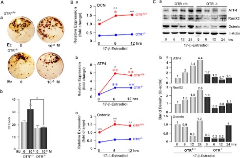
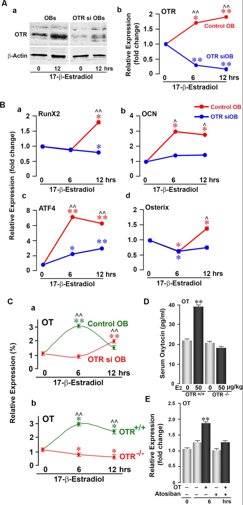
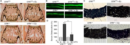
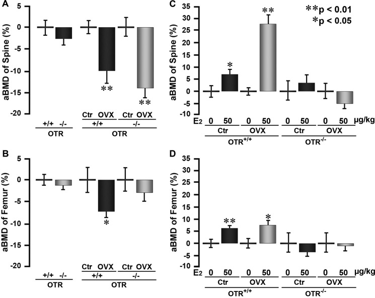
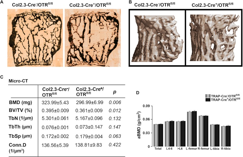
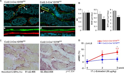
Estrogen uses two mechanisms to exert its effect on the skeleton: it inhibits bone resorption by osteoclasts and, at higher doses, can stimulate bone formation. Although the antiresorptive action of estrogen arises from the inhibition of the MAPK JNK, the mechanism of its effect on the osteoblast remains unclear. Here, we report that the anabolic action of estrogen in mice occurs, at least in part, through oxytocin (OT) produced by osteoblasts in bone marrow. We show that the absence of OT receptors (OTRs) in OTR(-/-) osteoblasts or attenuation of OTR expression in silenced cells inhibits estrogen-induced osteoblast differentiation, transcription factor up-regulation, and/or OT production in vitro. In vivo, OTR(-/-) mice, known to have a bone formation defect, fail to display increases in trabecular bone volume, cortical thickness, and bone formation in response to estrogen. Furthermore, osteoblast-specific Col2.3-Cre(+)/OTR(fl/fl) mice, but not TRAP-Cre(+)/OTR(fl/fl) mice, mimic the OTR(-/-) phenotype and also fail to respond to estrogen. These data attribute the phenotype of OTR deficiency to an osteoblastic rather than an osteoclastic defect. Physiologically, feed-forward OT release in bone marrow by a rising estrogen concentration may facilitate rapid skeletal recovery during the latter phases of lactation.


Figures

FIGURE 1.
FIGURE 1.
Estrogen-induced osteoblastogenesis is attenuated in OTR−/− cells. A, representative images (panel a) and colony counts (panel b) of ex vivo Von Kossa-positive mineralizing colonies (osteoblast colony-forming units (CFU-ob)) arising from wild-type (OTR+/+) or OTR−/− bone marrow stromal cells with either 17β-estradiol (E2; 10−8 m) or vehicle for 21 days. For panel b, zero dose versus 17β-estradiol (10−8 m) (*, p < 0.05) and wild-type versus OTR−/− (^, p < 0.05) (in duplicate). B, panel a, qPCR for osteocalcin (OCN) gene expression in OTR+/+ and OTR−/− osteoblasts for 0, 6, and 12 h. Expression was normalized to GAPDH and is plotted as -fold increase from the 0-h wild-type sample. Panels b and c, qPCR of wild-type (red) and OTR−/− (blue) calvarial osteoblasts showing changes in the expression of ATF4 (panel b) and Osterix (panel c) mRNA levels after treatment with 17β-estradiol (10−8 m) for 0, 6, or 12 h. Expression was normalized to GAPDH and plotted as -fold increase from the 0-h wild-type sample. Statistics were as follows: Student's t test with the Bonferroni correction, comparing 6- and/or 12-h time points each versus 0 h (*, p < 0.05; **, p < 0.01), as well as wild-type versus OTR−/− cells at all time points (^, p < 0.05; ^^, p < 0.01) (done twice in triplicate). C, panel a, OTR+/+ and OTR−/− calvarial osteoblasts were evaluated for changes in their expression of the osteoblast transcription factors ATF4, Runx2, and Osterix by Western blotting after treatment with 17β-estradiol (10−8 m) for 6, 12, and 24 h. Panel b, the band density was quantitated after normalizing to the loading control β-actin.
FIGURE 2.
FIGURE 2.
OTR knockdown attenuates estrogen-induced osteoblast differentiation. A, Western blotting (panel a) and qPCR (panel b) of MC3T3-E1 pre-osteoblasts (OBs) showing effective siRNA-mediated knockdown of the OTR. A 12-h time course was performed to demonstrate that OTR mRNA remained knocked down following 17β-estradiol (10−8 m) addition. B, qPCR of MC3T3-E1 pre-osteoblastic cells expressing either the OTR siRNA (OTR siOB) or empty vector (control OB) showing 17β-estradiol-induced changes in the expression of Runx2 (panel a), osteocalcin (OCN; panel b), ATF4 (panel c), and Osterix (panel d) mRNAs at 0, 6, and 12 h. Expression was normalized to GAPDH. C, effects of 17β-estradiol (10−8 m) on OT mRNA expression in OTR siRNA-silenced (OTR si OB; red) or vector-treated (Control OB; green) MC3T3-E1 cells (panel a) and in OTR−/− (red) or wild-type (green) osteoblasts (panel b) at 0, 6, and 12 h. mRNA expression is plotted as a percentage of 0 h and normalized to GAPDH. D, serum OT (by ELISA) in wild-type and OTR−/− mice after a 12-h treatment with either 17β-estradiol (E2; 50 μg/kg; black bars) or a control (gray bars). E, qPCR for OT mRNA showing that treatment of MC3T3-E1 cells with OT (10−8 m) increased OT mRNA at 6 h. To evaluate the specificity of OT-induced OT mRNA up-regulation, the OTR antagonist atosiban (10−8 m) was applied 20 min prior to OT addition. OT mRNA expression was normalized to GAPDH and is plotted as -fold increase from the 0-h non-treated sample. Statistics were as follows: Student's t test with the Bonferroni correction, comparing 6- and/or 12-h time points each versus 0 h (*, p < 0.05; **, p < 0.01), as well as wild-type versus OTR siRNA cells at all time points (^, p < 0.05; ^^, p < 0.01) (in triplicate). For D, comparisons between zero-dose and 17β-estradiol injections as described above (**, p < 0.01) (in duplicate).
FIGURE 3.
FIGURE 3.
Ability of estrogen to increase bone mass in wild-type mice requires functional OTRs. Two-month-old OTR−/− mice or wild-type (OTR+/+) littermates were treated with 17β-estradiol (E2; 50 μg/kg) or placebo biweekly (cumulative dose of 100 μg/kg/week). Representative images show the effect of 17β-estradiol on trabecular bone volume (A), double-labeled surfaces (B), and cortical thickness (C) in wild-type and OTR−/− mice. D, serum osteocalcin (by ELISA) 12 h after a single 17β-estradiol injection of 50 μg/kg. Statistics were as follows: Student's t test with the Bonferroni correction, comparing 17β-estradiol treatment versus zero dose (*, p < 0.05; **, p < 0.01), as well as wild-type versus OTR−/− mice (^^, p < 0.01).
FIGURE 4.
FIGURE 4.
Ability of estrogen to increase bone mass in ovariectomized mice is dependent on OTRs. Shown are areal BMD (aBMD; PIXImus) measurements at the spine and femur of sham-operated (control (Ctr)) and ovariectomized (OVX) OTR−/− and wild-type (OTR+/+) mice that were injected with 17β-estradiol (E2; 50 μg/kg) or placebo biweekly (cumulative dose of 100 μg/kg/week). A and B, effect of ovariectomy versus sham operation in the two genotypes. C and D, effect of 17β-estradiol versus placebo within the various groups. Statistics were as follows: means ± S.E. of the percent change shown using the respective controls (n = 5–6/group) and Student's t test with the Bonferroni correction, comparing ovariectomized versus control mice (A and B) and 17β-estradiol versus zero dose (C and D) (*, p < 0.05; **, p < 0.01).
FIGURE 5.
FIGURE 5.
Osteoblast-specific deletion of OTRs recapitulates global OTR deficiency. The skeletons of 16-week-old osteoblast-specific Col2.3-Cre+/OTRfl/fl mice or control Col2.3-Cre/OTRfl/fl littermates were phenotyped for differences in static and dynamic histomorphometry parameters. Representative images show von Kossa-stained trabecular bone (A) and micro-computed tomography of trabecular bone (B). C, micro-computed tomography (Micro-CT)-derived estimates of volumetric BMD, BV/TV, Tb.N, trabecular thickness (TbTh), trabecular spacing (TbSp), and connectivity density (Conn.D) in the two genotypes (units as shown). D, areal BMD (aBMD) using PIXImus of the osteoclast-specific TRAP-Cre+/OTRfl/fl mice or control TRAP-Cre/OTRfl/fl littermates recorded at the stated sites. Statistics were as follows: Student's t test with the Bonferroni correction, comparing Col2.3-Cre+/OTRfl/fl versus Col2.3-Cre/OTRfl/fl mice (C) (p values as shown) and TRAP-Cre+/OTRfl/fl versus TRAP-Cre/OTRfl/fl mice (D). L, left; R, right.
FIGURE 6.
FIGURE 6.
Osteoblast OTR mediates the anabolic action of estrogen on the skeleton. Representative images show double labeling with calcein (green) and xylenol orange (red) (see “Experimental Procedures”) (A) together with estimates of mineralizing surface (MS), MAR, and BFR (B) in Col2.3-Cre+/OTRfl/fl and Col2.3-Cre/OTRfl/fl mice. C, TRAP staining to show resorption areas, together with estimates of resorbed surface (Resorbed S./BPm.) in the two respective genotypes. D, effect of 17β-estradiol (50 μg/kg) or placebo biweekly (cumulative dose of 100 μg/kg/week) on areal BMD in Col2.3-Cre+/OTRfl/fl and Col2.3-Cre/OTRfl/fl (control) mice. Statistics were as follows: mean ± S.E. (n = 6–19/group) and Student's t test with the Bonferroni correction, comparing Col2.3-Cre+/OTRfl/fl versus Col2.3-Cre/OTRfl/fl mice (B and D) (^, p < 0.05; ^^, p < 0.01) and 17β-estradiol versus zero dose (D) (*, p < 0.05; **, p < 0.01).

References

    1. Breton C., Di Scala-Guenot D., Zingg H. H. (2001) Oxytocin receptor gene expression in rat mammary gland: structural characterization and regulation. J. Mol. Endocrinol. 27, 175–189 - PubMed
    1. Blanks A. M., Thornton S. (2003) The role of oxytocin in parturition. BJOG 110, 46–51 - PubMed
    1. Kovacs C. S. (2001) Calcium and bone metabolism in pregnancy and lactation. J. Clin. Endocrinol. Metab. 86, 2344–2348 - PubMed
    1. VanHouten J. N., Wysolmerski J. J. (2003) Low estrogen and high parathyroid hormone-related peptide levels contribute to accelerated bone resorption and bone loss in lactating mice. Endocrinology 144, 5521–5529 - PubMed
    1. Michalakis K., Peitsidis P., Ilias I. (2011) Pregnancy- and lactation-associated osteoporosis. Endocr. Regul. 45, 43–47 - PubMed

Publication types

MeSH terms