. 2012 Aug 17;287(34):28717-26.
doi: 10.1074/jbc.M112.383521. Epub 2012 Jul 2.

Long isoform mouse selenoprotein P (Sepp1) supplies rat myoblast L8 cells with selenium via endocytosis mediated by heparin binding properties and apolipoprotein E receptor-2 (ApoER2)

Affiliations

Long isoform mouse selenoprotein P (Sepp1) supplies rat myoblast L8 cells with selenium via endocytosis mediated by heparin binding properties and apolipoprotein E receptor-2 (ApoER2)

Suguru Kurokawa et al. J Biol Chem. .

Abstract

In vivo studies have shown that selenium is supplied to testis and brain by apoER2-mediated endocytosis of Sepp1. Although cultured cell lines have been shown to utilize selenium from Sepp1 added to the medium, the mechanism of uptake and utilization has not been characterized. Rat L8 myoblast cells were studied. They took up mouse Sepp1 from the medium and used its selenium to increase their glutathione peroxidase (Gpx) activity. L8 cells did not utilize selenium from Gpx3, the other plasma selenoprotein. Neither did they utilize it from Sepp1(Δ240-361), the isoform of Sepp1 that lacks the selenium-rich C-terminal domain. To identify Sepp1 receptors, a solubilized membrane fraction was passed over a Sepp1 column. The receptors apoER2 and Lrp1 were identified in the eluate by mass spectrometry. siRNA experiments showed that knockdown of apoER2, but not of Lrp1, inhibited (75)Se uptake from (75)Se-labeled Sepp1. The addition of protamine to the medium or treatment of the cells with chlorate also inhibited (75)Se uptake. Blockage of lysosome acidification did not inhibit uptake of Sepp1 but did prevent its digestion and thereby utilization of its selenium. These results indicate that L8 cells take up Sepp1 by an apoER2-mediated mechanism requiring binding to heparin sulfate proteoglycans. The presence of at least part of the selenium-rich C-terminal domain of Sepp1 is required for uptake. RT-PCR showed that mouse tissues express apoER2 in varying amounts. It is postulated that apoER2-mediated uptake of long isoform Sepp1 is responsible for selenium distribution to tissues throughout the body.


Figures

FIGURE 1.
FIGURE 1.
Uptake of 75Se from 75Se-labeled Sepp1 by L8 cells and inhibition of that uptake by serum selenoproteins. L8 cells were incubated in medium containing 2% Gpx3−/− mouse serum that contained 75Se-labeled Sepp1 as the only selenoprotein. A, the 75Se content of the cells was determined at intervals of up to 10 h of incubation. B, the effect of non-labeled Sepp1 on uptake of 75Se from 75Se-labeled Sepp1 was determined. 75Se uptake was determined in cells cultured for 5 h with Gpx3−/− mouse serum added to the medium in different concentrations. C, the effect of non-labeled Gpx3 and the shortest isoform of Sepp1 (Sepp1Δ240–361) on uptake of 75Se from 75Se-labeled Sepp1 was determined. 75Se uptake was determined in cells cultured for 5 h with Sepp1Δ240–361/Δ240–361 mouse serum added to the medium in different concentrations. All values are the means (n = 4) with 1 S.D. indicated by the brackets. Asterisks indicate values significantly different (p < 0.05) from the 30-min value (panel A) or the value with no unlabeled serum added (panels B and C) by Tukey's multiple comparison test applied after 1-way ANOVA of the groups.
FIGURE 2.
FIGURE 2.
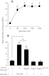
Response of L8 cell Gpx activity to the addition of different selenium sources to the medium. Selenium-depleted cells were cultured for 24 h with a selenium source and were then assayed for Gpx activity. A, selenite at different concentrations was the selenium source. B, selenium sources were 100 nm selenite and serum that contained single selenoproteins: Sepp1 (83 nm selenium), Gpx3 (98 nm selenium), and Sepp1Δ240–361 (101 nm selenium). The values shown are the means of three experiments carried out in duplicate with 1 S.D. indicated by the bracket. The asterisks in A indicate values significantly different (p < 0.05) from the value with no selenite added by Tukey's multiple comparison test applied after 1-way ANOVA of the groups and in B indicate values that were significantly different (p < 0.05) from the basal value by Student's t test.
FIGURE 3.
FIGURE 3.
Influence of heparin binding properties on L8 cell 75Se uptake from 75Se-labeled Sepp1. L8 cells were cultured for 5 h with 2% Gpx3−/− mouse serum that contained “all-isoform” 75Se-labeled Sepp1 as the only selenoprotein (panels A, B, and C) or 75Se-labeled selenite at 100 nm (panels D, E, and F). A and D, heparin was added to the medium at the concentrations indicated. B and E, protamine was added to the medium at the concentrations indicated. C and F, chlorate was added to the medium at the concentrations indicated 24 h before the 75Se source was introduced. Values shown are the means (n = 4) with 1 S.D. indicated by the bracket. Asterisks indicate values significantly different (p < 0.05) from the initial value by Tukey's multiple comparison test applied after one-way ANOVA of the groups.
FIGURE 4.
FIGURE 4.
Effect of knock down of receptors apoER2 and/or Lrp1 on 75Se uptake from 75Se-labeled Sepp1 by L8 and H9c2 cells. A, L8 cells were cultured for 48 h with siRNA, and then relative mRNA levels were determined. Values are the means (n = 3) with 1 S.D. indicated by the bracket. The asterisk indicates values different from control siRNA (p < 0.05) by Student's t test. B, uptake in 5-h incubations of 75Se by L8 cells with knockdown of receptors is shown. Values are the means of four experiments done in duplicate with 1 S.D. indicated by the bracket. Asterisks indicate that control was different (p < 0.05) from apoER2 and double knockdowns by Student's t test. C, H9c2 cells were cultured for 48 h with siRNA, and relative mRNA levels were determined. Values are the means (n = 3) with 1 S.D. indicated by the bracket. The asterisk indicates values different (p < 0.05) from control siRNA by Student's t test. D, shown is uptake in 5-h incubations of 75Se by H9c2 cells with knockdown of receptors. Values are the means of four experiments done in duplicate with 1 S.D. indicated by the bracket. The asterisk indicates that control is different (p < 0.05) from apoER2 and double knockdowns by Student's t test.
FIGURE 5.
FIGURE 5.
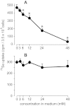
Effect of inhibiting clathrin and caveolin endocytosis pathways on the uptake of 75Se from 75Se-labeled Sepp1. L8 cells were incubated for 30 min in serum-free medium containing the indicated concentrations of chlorpromazine (A) or nystatin (B) before serum that contained 75Se-labeled Sepp1 was added. After 5 h, cells were washed, and 75Se was determined in them. Values are the means (n = 4) with 1 S.D. indicated by the bracket. Asterisks indicate values significantly different (p < 0.05) from the initial value by Tukey's multiple comparison test applied after one-way ANOVA of the groups.
FIGURE 6.
FIGURE 6.
Requirement of lysosomal function for L8 cell utilization of Sepp1 selenium. A, shown is the effect of 100 μm chloroquine on Gpx activity in L8 cells cultured for 24 h with Gpx3−/− serum containing Sepp1 (98 nm selenium) or with selenite (100 nm selenium). B, shown is the effect of 100 nm bafilomycin A1 on Gpx activity in L8 cells cultured with Gpx3−/− serum containing Sepp1 (98 nm selenium) or with selenite (100 nm selenium). C, 75Se uptake by L8 cells with bafilomycin A1 (100 nm) or chloroquine (100 μm) added to the medium is shown. Values in A–C are the means (n = 3 experiments carried out on duplicate plates) with 1 S.D. indicated by the bracket. Treated values were not significantly different from the control values (p < 0.05) by Student's t test. D, shown is the effect of chloroquine addition on Sepp1 form in L8 cells. L8 cells were incubated for 5 h with serum containing 75Se-labeled Sepp1 with or without 100 μm chloroquine. After washing, cells were lysed with SDS-PAGE buffer. Whole-cell lysates were subjected to SDS-PAGE and autoradiography (lanes 1 and 2) or to purification of Sepp1 using 9S4 antibody before the eluate was subjected to SDS-PAGE and autoradiography (lanes 3 and 4). Lane 5 shows an autoradiogram of 1 μl serum from a Gpx3−/− mouse that had been injected with 75Se-labeled selenite.
FIGURE 7.
FIGURE 7.
Relative abundance of apoER2 mRNA in tissues of mice fed diet supplemented with 0.25 mg of selenium/kg as selenite. Total RNA was extracted from designated tissues and used for cDNA synthesis and real-time PCR analysis. Levels of each tissue RNA are shown relative to hypoxanthine phosphoribosyltransferase. The brain value was set as 1, and the quantitation of each tissue is relative to it. Values are the means (n = 3) with the bracket indicating 1 S.D. The panel on the right shows tissues with very low relative mRNA expression (less than 3% of brain). TQ, relative quantitation.

References

    1. Hill K. E., Zhou J., McMahan W. J., Motley A. K., Atkins J. F., Gesteland R. F., Burk R. F. (2003) Deletion of selenoprotein P alters distribution of selenium in the mouse. J. Biol. Chem. 278, 13640–13646 - PubMed
    1. Schomburg L., Schweizer U., Holtmann B., Flohé L., Sendtner M., Köhrle J. (2003) Gene disruption discloses role of selenoprotein P in selenium delivery to target tissues. Biochem. J. 370, 397–402 - PMC - PubMed
    1. Burk R. F., Hill K. E. (2005) Selenoprotein P. An extracellular protein with unique physical characteristics and a role in selenium homeostasis. Annu. Rev. Nutr. 25, 215–235 - PubMed
    1. Burk R. F., Hill K. E. (2009) Selenoprotein P-Expression, functions, and roles in mammals. Biochim. Biophys. Acta 1790, 1441–1447 - PMC - PubMed
    1. Ma S., Hill K. E., Caprioli R. M., Burk R. F. (2002) Mass spectrometric characterization of full-length rat selenoprotein P and three isoforms shortened at the C terminus. Evidence that three UGA codons in the mRNA open reading frame have alternative functions of specifying selenocysteine insertion or translation termination. J. Biol. Chem. 277, 12749–12754 - PubMed

Publication types

MeSH terms

LinkOut - more resources