. 2012 Aug 10;287(33):27499-509.
doi: 10.1074/jbc.M111.323360. Epub 2012 Jul 2.

Cell adhesion-dependent serine 85 phosphorylation of paxillin modulates focal adhesion formation and haptotactic migration via association with the C-terminal tail domain of talin

Affiliations

Cell adhesion-dependent serine 85 phosphorylation of paxillin modulates focal adhesion formation and haptotactic migration via association with the C-terminal tail domain of talin

Tae Kyoung Kwak et al. J Biol Chem. .

Abstract

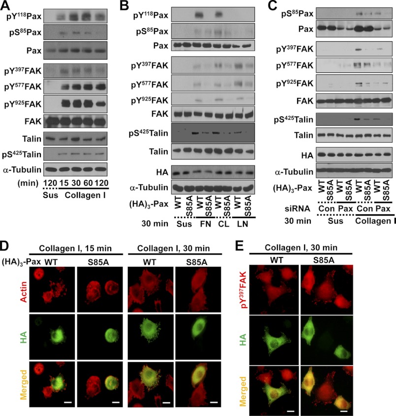
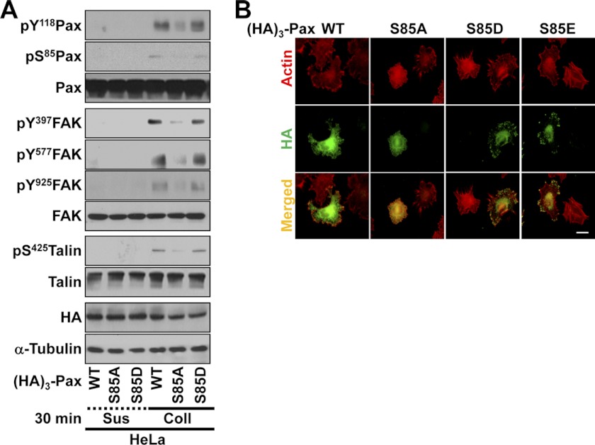
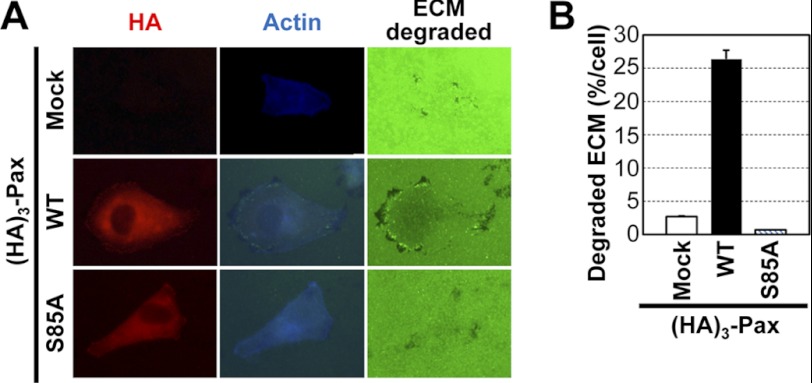
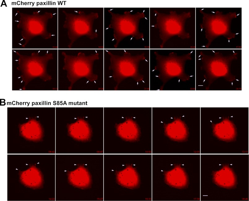
Integrin-mediated adhesion to extracellular matrix proteins is dynamically regulated during morphological changes and cell migration. Upon cell adhesion, protein-protein interactions among molecules at focal adhesions (FAs) play major roles in the regulation of cell morphogenesis and migration. Although tyrosine phosphorylation of paxillin is critically involved in adhesion-mediated signaling, the significance of paxillin phosphorylation at Ser-85 and the mechanism by which it regulates cell migration remain unclear. In this study, we examined how Ser-85 phosphorylation of paxillin affects FA formation and cell migration. We found that paxillin phosphorylation at Ser-85 occurred during HeLa cell adhesion to collagen I and was concomitant with tyrosine phosphorylation of both focal adhesion kinase and talin. However, the non-phosphorylatable S85A mutant of paxillin impaired cell spreading, FA turnover, and migration toward collagen I but not toward serum. Furthermore, whereas the (presumably indirect) interaction between paxillin and the C-terminal tail of talin led to dynamic FAs at the cell boundary, S85A paxillin did not bind talin and caused stabilized FAs in the central region of cells. Together, these observations suggest that cell adhesion-dependent Ser-85 phosphorylation of paxillin is important for its interaction with talin and regulation of dynamic FAs and cell migration.


Figures

FIGURE 1.
FIGURE 1.
Ser-85 phosphorylation of paxillin is dependent on cell adhesion. HeLa cells without (A) or with transfection with HA3-paxillin (paxillin or S85A) for 36 h (C, D, and E) were collected, rolled over at 37 °C for 1 h, and maintained in suspension (Sus) for 2 h. Cells were reseeded onto culture dishes newly precoated with collagen I (10 μg/ml) for the indicated times prior to harvesting and immunoblot analysis of whole cell lysates with the indicated antibodies (A–C) or reseeded on collagen I-precoated coverslips for 15 or 30 min before processing for indirect immunostaining or actin staining (D and E). Anti-HA tag antibody (green; D) or anti-HA tag (green) and anti-phospho-Tyr-397 FAK (pY397FAK; red) primary antibodies and the appropriate secondary antibodies (E) were used. Scale bars = 20 μm (D and E). B, HeLa cells transfected with HA3-paxillin (WT or S85A) for 36 h were kept in suspension or reseeded onto culture dishes precoated with 10 μg/ml fibronectin (FN), collagen I (CL), or laminin I (LN) for 30 min as described above. The cells were then harvested, and whole cell lysates were subjected to immunoblot analysis for the indicated molecules. The data represent three independent experiments. pS85Pax, phospho-Ser-85 paxillin; Con, control.
FIGURE 2.
FIGURE 2.
Paxillin Ser-85 phosphorylation affects cell adhesion signaling activity, FA formation, and actin organization. A, cells were transfected with HA3-paxillin (WT, S85A, or S85D) for 36 h before being kept in suspension (Sus) or reseeded onto collagen I (Coll; 10 μg/ml)-precoated culture dishes for 30 min. Whole cell lysates were prepared for standard Western blots for the indicated molecules. pS85Pax, phospho-Ser-85 paxillin. B, HeLa cells were transfected with HA3-paxillin (WT, S85A, S85D, or S85E) for 36 h before replating on collagen I (10 μg/ml)-precoated coverslips for 30 min. The cells were stained for actin using phalloidin and immunostained for HA3-paxillin using anti-HA antibody. Scale bar = 10 μm. The data shown represent three independent experiments.
FIGURE 3.
FIGURE 3.
Paxillin Ser-85 phosphorylation is important for haptotactic cell migration. A and B, cells were transfected with mock or HA3-paxillin (WT or S85A) constructs for 36 h before a Transwell migration assay, in which the lower chamber was filled with 1% BSA and DMEM-H (negative control), 10% FBS, or serum-free medium containing 10 μg/ml collagen I (Coll) for 12 h. After incubation, migrated cells on the chamber filter were stained with Diff-Quik solution (Medion Diagnostics), and at least five random images for each condition (A) were imaged to calculate means ± S.D. (B). C, cells were transfected with HA3-paxillin (WT or S85A) for 36 h and kept in suspension (Sus) or replated onto collagen I-precoated culture dishes for 30 min in the presence of 10% FBS before harvesting and analysis of whole cell lysates by immunoblotting for the indicated molecules. D and E, assay for cell migration through the Transwell for 6 h was performed as described for A. Cells were pretreated with SB202190 (SB) 30 min before loading cells into the upper chambers. Migrated cells from representative images (D) were calculated for means ± S.D. (E). F, cells were transfected and manipulated as described for C in the absence (−) or presence of SB202190 pretreatment before harvesting and analysis of whole cell lysates by immunoblotting for the indicated molecules. The data shown represent three independent experiments. pS85Pax, phospho-Ser-85 paxillin.
FIGURE 4.
FIGURE 4.
Paxillin Ser-85 phosphorylation is important for invasive protrusion formation. Cells were either mock-transfected or transfected with HA3-paxillin (HA3-Pax; WT or S85A) for 36 h before reculturing on Oregon Green® 488-conjugated gelatin for 6 h. ECM degradation by invasive protrusions along the cell boundary was confirmed by actin staining concurrent with detection of black spots (degradation of Oregon Green 488-conjugated gelatin) (A). The numbers of cells with degraded ECM were counted among at least 100 cells for each condition, and the percentage of the ECM-degraded area out of the total area was calculated to obtain means ± S.D. from three different experiments (B).
FIGURE 5.
FIGURE 5.
WT (but not S85A) paxillin binds to talin. A, immunoprecipitates (IP) using anti-HA antibody from extracts of mock-transfected or HA3-paxillin (HA3-Pax; WT or S85A)-transfected cells were run in a gel and silver-stained. Arrowheads indicate certain proteins immunoprecipitated from WT paxillin-transfected cell extracts but not from mock-transfected or S85A (SA) paxillin-transfected cell extracts. IgG-h and IgG-l, IgG heavy and light chains, respectively. B, recombinant GST alone or GST-paxillin (WT or S85A) proteins bound to glutathione-agarose beads were mixed with whole cell lysates (WCL) from HeLa cells overnight at 4 °C, and the pulldown samples were analyzed by Western blotting using anti-GST or anti-talin antibody. C, HeLa cells were transfected with HA3-paxillin (WT or S85A) for 36 h and kept in suspension (Sus) or reseeded onto collagen I (Coll)-precoated culture dishes for 30 min prior to harvesting. Whole cell lysates were immunoprecipitated with anti-HA antibody as described under “Experimental Procedures.” The immunoprecipitates and whole cell lysates were analyzed in parallel by standard Western blotting with anti-HA, anti-FAK, or anti-talin antibody. D–F, HeLa cells were cotransfected with HA3-paxillin (WT or S85A) and the GFP-tagged N-terminal head (Head) or C-terminal tail (C-tail) domain of talin for 36 h. The cells were then kept in suspension or reseeded onto collagen I-precoated culture dishes for 30 min prior to harvesting. Whole cell lysates were subjected to standard Western blot (WB) analysis using the indicated antibodies (D) or co-immunoprecipitation using anti-GFP (E) or anti-HA (F) antibody prior to immunoblotting in parallel with lysates (Input) using antibodies for the indicated molecules. pS85Pax, phospho-Ser-85 paxillin. G, recombinant GST-talin tail fragments (D1–D4) on beads were mixed with HeLa cell extracts as described under “Experimental Procedures.” The proteins on beads were subjected to standard Western blotting for paxillin or GST. H, cells were mock-transfected or transfected with HA3-paxillin (WT or S85A) for 36 h before manipulating cells as described for C. Immunoprecipitates with anti-HA antibody were immunoblotted in parallel with lysates for the indicated molecules. The data are representative of three independent experiments.
FIGURE 6.
FIGURE 6.
FA formation is regulated by WT (but not S85A) paxillin together with the talin C-terminal tail domain. Cells were cotransfected with HA3-paxillin (HA3-Pax; WT or S85A) and GFP-WT talin (A), the N-terminal head domain (B), or the C-terminal tail domain (C and D) for 36 h. Alternatively, cells were transiently transfected with control siRNA (siControl) or siRNA against paxillin (siPaxillin) (E) or cotransfected with paxillin siRNA and HA3-S85A paxillin (F) for 36 h. The cells were then replated onto collagen I (10 μg/ml)-precoated coverslips for 30 min before staining with anti-HA3 tag (red, A–C; and green, F), anti-actin (red, E), anti-paxillin (green, E), or anti-vinculin (red, D and F) antibody. Scale bars = 10 μm (A–D and F) and 20 μm (E). The data shown represent three independent experiments.
FIGURE 7.
FIGURE 7.
Time-lapse images of mCherry-paxillin (WT or S85A)-expressing cells. Cells were transiently transfected with mCherry-tagged WT (A) or S85A mutant (B) paxillin for 36 h and replated onto collagen I (2 μg/ml)-precoated coverslips for 30 min within 0.2% BSA and DMEM-H before analysis with time-lapse microscopy. Images were saved over 30 min, and snap pictures in series over at least 10 min are represented. A, in images of mCherry-WT paxillin-transfected cells, dynamic turnover of paxillin-positive spots (arrows) is evidenced by newly formed spots compared with the immediately preceding snap picture. B, in images of mCherry-S85A paxillin-transfected cells, the positive staining spots (arrowheads) were less dynamic, and the staining was sustained for longer times. Images shown represent cells from several analyses (mCherry-WT paxillin, n = 9; and mCherry-S85A paxillin, n = 11).

Similar articles

Cited by

References

    1. Danen E. H. (2009) Integrin proteomes reveal a new guide for cell motility. Sci. Signal. 2, pe58. - PubMed
    1. Mitra S. K., Schlaepfer D. D. (2006) Integrin-regulated FAK-Src signaling in normal and cancer cells. Curr. Opin. Cell Biol. 18, 516–523 - PubMed
    1. Schaller M. D. (2010) Cellular functions of FAK kinases: insight into molecular mechanisms and novel functions. J. Cell Sci. 123, 1007–1013 - PubMed
    1. Lock J. G., Wehrle-Haller B., Stromblad S. (2008) Cell-matrix adhesion complexes: master control machinery of cell migration. Semin. Cancer Biol. 18, 65–76 - PubMed
    1. Carragher N. O., Frame M. C. (2004) Focal adhesion and actin dynamics: a place where kinases and proteases meet to promote invasion. Trends Cell Biol. 14, 241–249 - PubMed

Publication types