. 2012 Aug 24;287(35):29672-8.
doi: 10.1074/jbc.M111.322404. Epub 2012 Jul 3.

Sequestosome 1/p62, a scaffolding protein, is a newly identified partner of IRS-1 protein

Affiliations

Sequestosome 1/p62, a scaffolding protein, is a newly identified partner of IRS-1 protein

Thangiah Geetha et al. J Biol Chem. .

Abstract

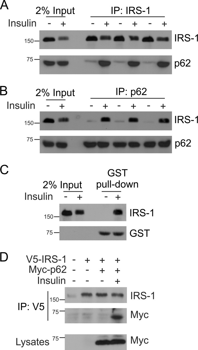
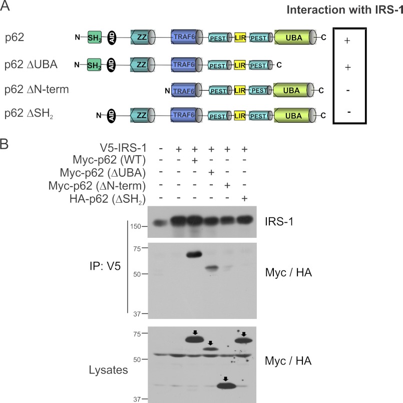
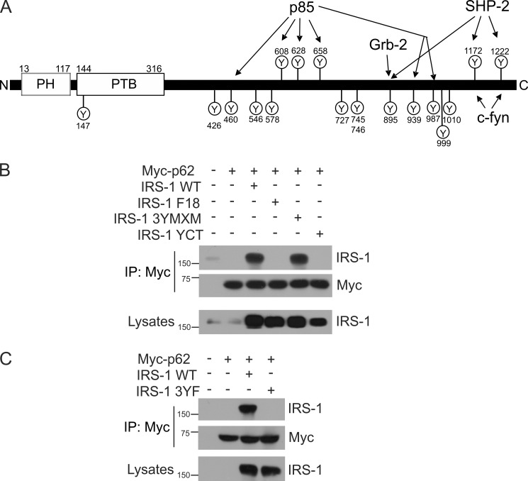
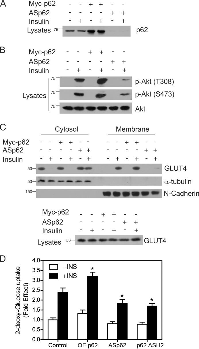
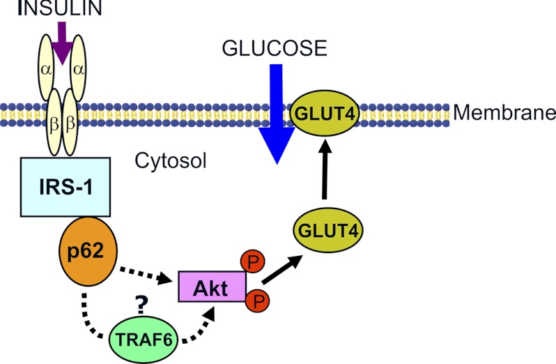
Defects in the insulin-signaling pathway may lead to the development of skeletal muscle insulin resistance, which is one of the earliest abnormalities detected in individuals with the metabolic syndrome and predisposes them to develop type 2 diabetes. Previous studies have shown that deletion of the mouse sequestosome 1/p62 gene results in mature-onset obesity that progresses to insulin and leptin resistance and, ultimately, type 2 diabetes. Sequestosome 1/p62 is involved in receptor-mediated signal transduction and functions as an intracellular signal modulator or adaptor protein. Insulin receptor substrate-1 (IRS-1) plays a central role in transducing the insulin signal via phosphorylation, protein-protein interactions, and protein modifications. Mapping studies demonstrated that the SH(2) domain at the amino terminus of sequestosome 1/p62 interacts with IRS-1 upon insulin stimulation. Further, IRS-1 interacts with p62 through its YMXM motifs at Tyr-608, Tyr-628, and/or Tyr-658 in a manner similar to its interaction with p85 of phosphoinositol 3-kinase. Overexpression of p62 increased phosphorylation of Akt, GLUT4 translocation, and glucose uptake, providing evidence that p62 participates in the insulin-signaling pathway through its interactions with IRS-1.


Figures

FIGURE 1.
FIGURE 1.
Insulin-dependent interaction between p62 and IRS-1. A, L6 myotubes were serum-starved for 4 h and either left untreated or stimulated with insulin (100 nm) for 15 min at 37 °C. One mg of lysate protein was immunoprecipitated (IP) with IRS-1 antibody and Western blotted with antibodies against IRS-1 or p62, n = 3. The lanes labeled Input indicate crude lysate. B, the above lysates were also immunoprecipitated with anti-p62 and Western blotted with IRS-1 and p62 antibody. C, L6 myotubes (400 μg) stimulated with or without insulin were interacted with an equivalent amount of GST-p62 in a pulldown assay. The association of IRS-1 was determined by Western blotting with IRS-1 and GST antibody. D, CHO/IR cells were transfected with V5-IRS-1 and Myc-p62 and stimulated with or without insulin for 15 min. The cells were lysed, immunoprecipitated with V5 antibody, and immunoblotted with IRS-1 and Myc antibody. Lysates were Western blotted with anti-Myc to assess the expression of the p62 plasmid. Experiments were replicated three times with similar results.
FIGURE 2.
FIGURE 2.
p62 interacts with IRS-1 through its SH2 domain. A, domain structures of full-length p62 and p62 mutants (ΔUBA, ΔN-term, and ΔSH2) were used to map the interaction with IRS-1. N, N terminus; AID, acidic interaction domain; LIR, LC3-interacting region; C, C terminus. B, CHO/IR cells were transfected with IRS-1 and wild-type Myc-tagged p62, ΔUBA, ΔN-term, or HA-p62ΔSH2 domain. The cells were stimulated with insulin (100 nm) for 15 min and lysed in Triton lysis buffer. The cell lysates were immunoprecipitated (IP) with anti-V5 followed by Western blotting with anti-IRS-1 and anti-Myc/HA. The lysates were also blotted with anti-Myc/HA to verify the expression of the p62 constructs. Experiments were replicated three times with similar results.
FIGURE 3.
FIGURE 3.
IRS-1 interacts with p62 through its YXXM motif. A, schematic representation of IRS-1 showing the various tyrosine residues. N, N terminus; PH, pleckstrin homology domain; PTB, phosphotyrosine binding domain; C, C terminus. B, CHO/IR cells were expressed with wild-type IRS-1 and the mutants F18, 3YMXM, or YCT along with Myc-tagged p62. The cells were serum-starved and stimulated with insulin (100 nm) for 15 min. p62 was immunoprecipitated (IP) using Myc antibody and Western blotted for IRS-1 and p62. C, CHO/IR cells were transfected with Myc-p62 along with wild-type IRS-1 or 3YF mutant and treated with insulin for 15 min. The co-interaction of p62 and IRS-1 was determined by immunoprecipitation with anti-Myc and Western blotting for anti-IRS-1 and anti-Myc. The lysates were Western blotted with anti-IRS-1 to verify the expression of the constructs. Experiments were replicated three times with similar results.
FIGURE 4.
FIGURE 4.
p62 influences insulin receptor signaling. A, CHO/IR cells were transfected with either Myc-tagged p62 or ASp62. The transfected cells were treated with or without insulin (100 nm) for 15 min at 37 °C. Lysates from control and transfected cells were Western blotted with p62. B, the above lysates (A) were immunoblotted for phospho-Akt Thr-308 (p-Akt (T308)) and phospho-Akt Ser-473 (p-Akt (S473)) and total Akt (Akt). C, the transfected cells from above (A) were incubated in the presence or absence of insulin for 20 min, fractionated into membrane and cytosolic fractions, and Western blotted with GLUT4, α-tubulin, and N-cadherin. The lysates were immunoblotted for total GLUT4. D, OEp62, ASp62, or p62ΔSH2 mutants were expressed in CHO/IR cells, cells were stimulated in the presence or absence of 100 nm insulin for 20 min, and the rates of 2-deoxy-d-[3H]glucose uptake were determined. Each bar in the graph indicates the -fold change relative to the control cells unstimulated with insulin, which was taken to be 1. Differences from the control value treated with insulin and other groups are statistically significant (*, p < 0.001). Error bars indicate S.D.
FIGURE 5.
FIGURE 5.
Model depicting the role of p62 in insulin signaling. p62 interacts with IRS-1 on insulin stimulation and leads to Akt activation, GLUT4 translocation, and glucose uptake. Effect of p62 on Akt phosphorylation (indicated by circled P) may involve other proteins such as TRAF6 (35, 38).

Similar articles

Cited by

References

    1. Bouzakri K., Koistinen H. A., Zierath J. R. (2005) Molecular mechanisms of skeletal muscle insulin resistance in type 2 diabetes. Curr. Diabetes Rev. 1, 167–174 - PubMed
    1. Lee J., Pilch P. F., Shoelson S. E., Scarlata S. F. (1997) Conformational changes of the insulin receptor upon insulin binding and activation as monitored by fluorescence spectroscopy. Biochemistry 36, 2701–2708 - PubMed
    1. Myers M. G., Jr., Grammer T. C., Brooks J., Glasheen E. M., Wang L. M., Sun X. J., Blenis J., Pierce J. H., White M. F. (1995) The pleckstrin homology domain in insulin receptor substrate-1 sensitizes insulin signaling. J. Biol. Chem. 270, 11715–11718 - PubMed
    1. Myers M. G., Jr., White M. F. (1996) Insulin signal transduction and the IRS proteins. Annu. Rev. Pharmacol. Toxicol. 36, 615–658 - PubMed
    1. Craparo A., O'Neill T. J., Gustafson T. A. (1995) Non-SH2 domains within insulin receptor substrate-1 and SHC mediate their phosphotyrosine-dependent interaction with the NPEY motif of the insulin-like growth factor I receptor. J. Biol. Chem. 270, 15639–15643 - PubMed

Publication types

MeSH terms