Clinical Trial
. 2012 Aug 17;287(34):28435-44.
doi: 10.1074/jbc.M112.385823. Epub 2012 Jul 3.

Polyphosphate is a novel pro-inflammatory regulator of mast cells and is located in acidocalcisomes

Affiliations
Clinical Trial

Polyphosphate is a novel pro-inflammatory regulator of mast cells and is located in acidocalcisomes

David Moreno-Sanchez et al. J Biol Chem. .

Abstract

Polyphosphate (polyP) is a pro-inflammatory agent and a potent modulator of the human blood-clotting system. The presence of polyP of 60 phosphate units was identified in rat basophilic leukemia (RBL-2H3) mast cells using specific enzymatic assays, urea-polyacrylamide gel electrophoresis of cell extracts, and staining of cells with 4,6-diamidino-2-phenylindole (DAPI), and the polyP-binding domain of Escherichia coli exopolyphosphatase. PolyP co-localizes with serotonin- but not with histamine-containing granules. PolyP levels greatly decreased in mast cells stimulated to degranulate by IgE. Mast cell granules were isolated and found to be acidic and decrease their polyP content upon alkalinization. In agreement with these results, when RBL-2H3 mast cells were loaded with the fluorescent calcium indicator fura-2 acetoxymethyl ester to measure their intracellular Ca(2+) concentration ([Ca(2+)](i)), they were shown to possess a significant amount of Ca(2+) stored in an acidic compartment different from lysosomes. PolyP derived from RBL-2H3 mast cells stimulated bradykinin formation, and it was also detected in human basophils. All of these characteristics of mast cell granules, together with their known elemental composition, and high density, are similar to those of acidocalcisomes. The results suggest that mast cells polyP could be an important mediator of their pro-inflammatory and pro-coagulant activities.


Figures

FIGURE 1.
FIGURE 1.
Detection of polyP in mast cells. A, epifluorescence microscopy with specific filters for localization of polyP (red) and DNA (blue). B, confocal fluorescence microscopy of fixed RBL-2H3 cells labeled with DAPI, showing localization of DNA (blue) and polyP (green). Fluorescent (left) and merged bright field (BF, right) images are shown. C, flow cytometry analysis of RBL-2H3 cells in the absence (Control) and presence of DAPI (+DAPI). The geometric mean fluorescence intensities for DAPI-polyP complexes (in the FL3 channel of the flow cytometer) are indicated. D, localization of polyP using the recombinant PPBD of E. coli PPX linked with an Xpress epitope tag. Samples were analyzed by confocal fluorescence microscopy as described under “Experimental Procedures.” Fluorescent (left) and its corresponding merge (center), and bright field (BF, right) images, are shown. A–D show representative experiments (n = 4). Scale bars, 10 μm (A, B, and D).
FIGURE 2.
FIGURE 2.
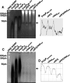
Urea-PAGE analysis of polyP extracted from mast cells and mast cell granules. A, polyP extracted from RBL-2H3 cells was electrophoresed by 6% urea-PAGE and stained with DAPI (negative staining). Chain lengths of commercial standards (polyP25, polyP45, and polyP65) are on the left. An arrow on the right shows the position of migration of RBL-2H3 sample (polyPRBL-2H3). In addition, extracted mast cell polyP was treated with an excess of ScPPX1 (PPX) and/or heparinase III (Hepase). Left lane is a DNA ladder. Note that DAPI bound to DNA did not photobleach. DNA remained positively stained whereas polyP was negatively stained. B, densitometric analysis of the labeled gel shown in A was performed at the zone where mast cell polyP was found (arrow). Analysis was performed using ImageJ 1.43u software. (C) PolyP extracted from RBL-2H3 isolated granules was electrophoresed and treated as described in A. An arrow on the right shows the position of migration of polyP granule sample (PolyPgranules). D, densitometric analysis of the labeled gel shown in A was performed at the zone where mast cell polyP was found (arrow). Analysis was performed as above. A–D show representative experiments (n = 3).
FIGURE 3.
FIGURE 3.
Localization of polyP, serotonin, and histamine in mast cells. PolyP co-localizes with serotonin (A) but not with histamine (B) in RBL-2H3 cells whereas serotonin only partially co-localizes with histamine (C), as shown by confocal immunofluorescence analysis. Fixed RBL-2H3 cells were labeled with the recombinant PPBD of E. coli PPX linked with an Xpress epitope tag as shown in Fig. 1D, and with specific antibodies for serotonin and histamine as described under “Experimental Procedures.” For each combination, independent fluorescent signals and their merged images are shown. Scale bars, 10 μm.
FIGURE 4.
FIGURE 4.
Decrease in intracellular polyP after mast cell activation. RBL-2H3 cells were stimulated to degranulate by sequential exposure to anti-DNP-IgE and DNP-BSA, as described under “Experimental Procedures.” A, confocal fluorescence microscopy of fixed cells before (Basal) and after activation (Activated), and labeled with DAPI to show polyP localization. Corresponding bright field images are shown in the right panels. A representative experiment is shown (n = 3). B, densitometric analysis of polyP labeling of 50 cells before (Basal) and after (Activ.) mast cell activation. Analysis was performed using ImageJ 1.43u software. Results show the mean ± S.D. (error bars) for three experiments. C, percentage of β-hexosaminidase activity in the supernatant compared with the total enzyme activity in the cell pellets before (Basal) and after (Activ.) mast cell activation. Results show the mean ± S.D. of 3 experiments.
FIGURE 5.
FIGURE 5.
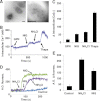
Mast cells have electron-dense, acidic, and calcium-rich organelles. A, electron microscopy of whole intact RBL-2H3 cells allowed to adhere to a Formvar- and carbon-coated grid and observed by transmission electron microscopy. The pictures show a detail of the intracellular granules with intragranular regions of high density. Scale bars, 0.2 μm. B, intracellular calcium measurements. RBL-2H3 cells were loaded with fura-2/AM and suspended in a medium with 100 μm EGTA as described under “Experimental Procedures.” At the times indicated, 40 μm GPN, 5 μm nigericin (NIG), 20 mm NH4Cl, and 1 μm thapsigargin (Thaps) were added. C, histograms summarizing the changes in [Ca2+]i (n = 3). D, changes in acridine orange fluorescence. RBL-2H3 cells were incubated with acridine orange, and their plasma membrane was permeabilized with digitonin, as described under “Experimental Procedures.” At the times indicated, 5 μm nigericin and 20 mm NH4Cl were added. E, histograms summarizing the changes in acridine orange fluorescence (n = 3).
FIGURE 6.
FIGURE 6.
PolyP content, acidity, and depletion of polyP after alkalinization, of isolated mast cell granules. A, short chain (SC) and long chain (LC) polyP in whole RBL-2H3 cells (cells) and isolated mast cell granules (granules). Results are the mean ± S.D. (error bars; n = 4). B, isolated mast cell granules incubated with acridine orange (A.O.) and analyzed by fluorescence microscopy as described under “Experimental Procedures.” BF, bright field image. Scale bars, 5 μm. C, short and long chain polyP levels were measured in isolated mast cell granules at different times after the addition of 10 μm nigericin (NIG) or 20 mm NH4Cl. Results are the mean ± S.D. of n = 4.
FIGURE 7.
FIGURE 7.
Human basophils possess polyP, and mast cell polyP induces bradykinin release in plasma. A, confocal fluorescence microscopy of human peripheral blood mononuclear cells labeled with specific anti-IgE FITC antibodies (IgE) and the recombinant PPBD of E. coli exopolyphosphatase (PPBD-PolyP) as described under “Experimental Procedures.” Independent fluorescent signals and their merged images are shown. Scale bar, 10 μm. B, flow cytometry analysis of whole fixed human blood cells after labeling with anti-IgE phycoerythrin antibodies. Dot plot represents a typical pattern of expression of IgE in human blood cells, in which basophilic granulocytes subset (Bas) and other blood cells (“IgEneg”) are indicated. The measurement of a representative sample is shown (n = 4). C, fixed samples of labeled blood cells, as described in A, were incubated in the absence (Control) or presence of DAPI (+DAPI). Cell fluorescence was determined on gated basophils (Bas, upper panel), and other blood cells (IgEneg, lower panel), in the FL3 channel that is specific for DAPI-polyP complexes (n = 4). D, differences in the geometric mean fluorescence intensity in the FL3 channel, after addition of DAPI in basophils (Bas), and other blood cells (IgEneg). Results show the mean ± S.D. (error bars) of the measurements made in samples from four individuals. Asterisk indicates significant differences, as determined by the Student's t test (p < 0.05). E, bradykinin release was quantified by ELISA in human plasma after incubation with synthetic polyP (polyP65, 10 μg/ml) or with polyP extracted from RBL-2H3 cells (polyPRBL-2H3). Results show the mean ± S.D. of measurements made in samples from three individuals.

References

    1. Morrissey J. H., Choi S. H., Smith S. A. (2012) Polyphosphate: an ancient molecule that links platelets, coagulation and inflammation. Blood 119, 5972–5979 - PMC - PubMed
    1. Rao N. N., Gómez-García M. R., Kornberg A. (2009) Inorganic polyphosphate: essential for growth and survival. Annu. Rev. Biochem. 78, 605–647 - PubMed
    1. Docampo R., Moreno S. N. (2011) Acidocalcisomes. Cell Calcium 50, 113–119 - PMC - PubMed
    1. Ruiz F. A., Lea C. R., Oldfield E., Docampo R. (2004) Human platelet dense granules contain polyphosphate and are similar to acidocalcisomes of bacteria and unicellular eukaryotes. J. Biol. Chem. 279, 44250–44257 - PubMed
    1. Müller F., Mutch N. J., Schenk W. A., Smith S. A., Esterl L., Spronk H. M., Schmidbauer S., Gahl W. A., Morrissey J. H., Renné T. (2009) Platelet polyphosphates are proinflammatory and procoagulant mediators in vivo. Cell 139, 1143–1156 - PMC - PubMed

Publication types

LinkOut - more resources