. 2012 Aug 17;287(34):28206-14.
doi: 10.1074/jbc.M112.384594. Epub 2012 Jul 3.

Chemically programmed bispecific antibodies that recruit and activate T cells

Affiliations

Chemically programmed bispecific antibodies that recruit and activate T cells

Huiting Cui et al. J Biol Chem. .

Abstract

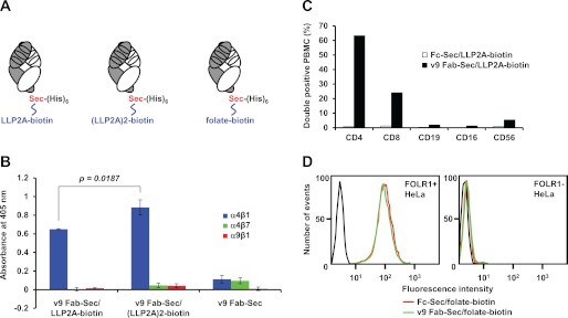
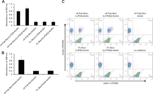
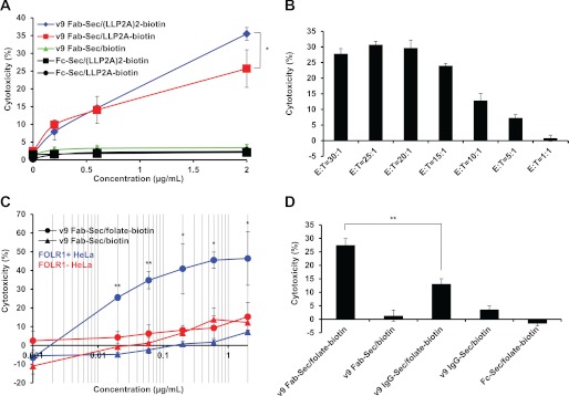
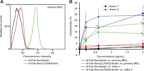
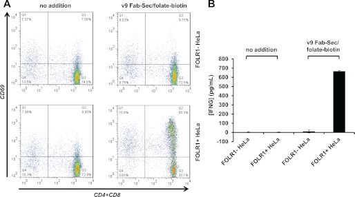
Bispecific antibodies (biAbs) that mediate cytotoxicity by recruiting and activating endogenous immune cells are an emerging class of next-generation antibody therapeutics. Of particular interest are biAbs of relatively small size (∼50 kDa) that can redirect cytotoxic T cells through simultaneous binding of tumor cells. Here we describe a conceptually unique class of biAbs in which the tumor cell specificity of a humanized antibody fragment that recognizes CD3 on T cells is chemically programmed through a C-terminal selenocysteine (Sec) residue. We demonstrate that through chemically programmed specificity for integrin α(4)β(1) or folate receptor 1 (FOLR1), and common specificity for CD3, these hybrid molecules exert potent and specific in vitro and ex vivo cytotoxicity toward tumor cell lines and primary tumor cells in the presence of primary T cells. Importantly, the generic nature of chemical programming allows one to apply our approach to virtually any specificity, promising a broad utility of chemically programmed biAbs in cancer therapy.


Figures

FIGURE 1.
FIGURE 1.
Concept and specificity of chemically programmed biAbs. A, drawing of anti-human CD3 Fab v9 with a Sec at the C-terminus of the heavy chain fragment followed by a hexa-histidine tag and chemically programmed with LLP2A-biotin (left panel), (LLP2A)2-biotin (center panel) for human integrin α4β1 binding, or folate-biotin (right panel) for human FOLR1 binding. The light chain is shown in white and the heavy chain fragment in gray. Each consists of one variable and one constant Ig domain. Also indicated are the three complementarity determining regions of each variable domain that mediate CD3 binding. B, binding of v9 Fab-Sec before (right) or after chemical programming with LLP2A-biotin (left) or (LLP2A)2-biotin (center) to immobilized human integrins α4β1 (blue), α4β7 (green), and α9β1 (red) in the presence of 1 mm MnCl2 as detected with HRP-conjugated goat anti-human κ light chain polyclonal antibodies (pAbs). Shown are mean values of triplicates ± S.D.. An unpaired two-tailed Student's t test was used to calculate p. C, binding of v9 Fab-Sec/LLP2A-biotin (black) detected with PE-conjugated streptavidin to normal human PBMC subpopulations gated with APC-conjugated mouse anti-human CD4, CD8, CD19, CD16, and CD56 mAbs. Shown is the percentage of cells in the double positive quadrant. Fc-Sec/LLP2A-biotin (white) was used as negative control. D, flow cytometry analysis of the binding of v9 Fab-Sec/folate-biotin (green), Fc-Sec/folate-biotin (red), or the detecting reagent PE-conjugated streptavidin alone (black) to FOLR1+ (left panel) and FOLR1- (right panel) HeLa cells.
FIGURE 2.
FIGURE 2.
Bispecificity of chemically programmed biAbs. A, adherence of CD3+ Jurkat cells incubated with 10 μg/ml bispecific conjugates (v9 Fab-Sec/LLP2A-biotin and v9 Fab-Sec/(LLP2A)2-biotin) and control conjugates (v9 Fab-Sec/biotin, Fc-Sec/LLP2A-biotin, and Fc-Sec/(LLP2A)2-biotin) to immobilized human integrin α4β1 in the presence of 1 mm MnCl2. B, adherence of CD3+ Jurkat cells incubated with 10 μg/ml bispecific conjugate (v9 Fab-Sec/folate-biotin) and control conjugates (v9 Fab-Sec/biotin and Fc-Sec/folate) to immobilized human FOLR1. In both experiments, the relative number of adherent cells was determined through a colorimetric lactate dehydrogenase assay. Shown are mean values of triplicates ± S.D. after subtraction of the background measured in the absence of conjugates. C, cell-cell association of CD3+ Jurkat cells loaded with red fluorescent dye PKH26 and integrin α4β1+ JeKo-1 cells loaded with green fluorescent dye CFSE. Cells were mixed in a 1:1 ratio and incubated for 30 min at room temperature in the absence or presence of 2 μg/ml bispecific conjugates (v9 Fab-Sec/LLP2A-biotin and v9 Fab-Sec/(LLP2A)2-biotin) and control conjugates (v9 Fab-Sec/biotin, Fc-Sec/LLP2A-biotin, and Fc-Sec/(LLP2A)2-biotin). Following resuspension in 1% (w/v) paraformaldehyde in PBS, double fluorescent Jurkat-JeKo-1 cell-cell associations were detected by flow cytometry (upper right quadrant).
FIGURE 3.
FIGURE 3.
Activity of chemically programmed biAbs. A, cytotoxicity of different concentrations of bispecific conjugates v9 Fab-Sec/(LLP2A)2-biotin (blue ♦) and v9 Fab-Sec/LLP2A-biotin (red ■) and control conjugates v9 Fab-Sec/biotin (green ▴), Fc-Sec/(LLP2A)2-biotin (black ■), and Fc-Sec/LLP2A-biotin (black ●), with normal human PBMC as effector cells (E) and JeKo-1 cells as target cells (T) at an E:T ratio of 20:1. B, cytotoxicity of 2 μg/ml v9 Fab-Sec/(LLP2A)2-biotin at the indicated seven E:T ratios. C, cytotoxicity of different concentrations of bispecific conjugate (v9 Fab-Sec/folate-biotin (●)) and control conjugate (v9 Fab-Sec/biotin (▴)) with normal human PBMC (E) and FOLR1+ (blue) and FOLR1- (red) HeLa cells (T) at an E:T ratio of 20:1. To support the log conversion, 0 μg/ml concentrations are shown as 0.001 μg/ml. D, comparison of the cytotoxicity of equimolar concentrations (4 nm) of v9 Fab-Sec/folate-biotin (0.2 μg/ml) and v9 IgG-Sec/folate-biotin (0.6 μg/ml) and control conjugates (v9 Fab-Sec/biotin (0.2 μg/ml), v9 IgG-Sec/biotin (0.6 μg/ml), and Fc-Sec/folate-biotin (0.2 μg/ml)) with normal human PBMC (E) and FOLR1+ HeLa cells (T) at an E:T ratio of 20:1. In all experiments, cytotoxicity was measured with the CytoTox-Glo cytotoxicity assay after a 16-h incubation at 37 °C. Shown are mean values of triplicates ± S.D.. An unpaired two-tailed Student's t test was used to calculate p. *, p < 0.05; **, p < 0.01, respectively. Actual p values were 0.0362 (A); from left to right, 0.0009, 0.0008, 0.0237, 0.0011, 0.0219 (C); and 0.0008 (D).
FIGURE 4.
FIGURE 4.
Primary cell binding and cytotoxicity of chemically programmed biAbs. A, flow cytometry analysis of the binding of v9 Fab-Sec/LLP2A-biotin (green), v9 Fab-Sec/biotin (red), or the detecting reagent PE-conjugated streptavidin alone (black) to primary MCL cells from a lymph node biopsy. B, cytotoxicity of different concentrations of bispecific conjugate v9 Fab-Sec/(LLP2A)2-biotin toward primary MCL cells (green) and JeKo-1 cells (blue) as target cells (T) in the presence of normal human PBMC from donor 1 (●) and donor 2 (▴) as effector cells (E). The cytotoxicity of corresponding concentrations of control conjugate v9 Fab-Sec/biotin toward primary MCL cells (red) and JeKo-1 cells (black) was determined in parallel. In all experiments, cytotoxicity was measured with the CytoTox-Glo cytotoxicity assay after a 16-h incubation at 37 °C and an E:T ratio of 20:1. Shown are mean values of triplicates ± S.D. For the cytotoxicity of v9 Fab-Sec/(LLP2A)2-biotin versus v9 Fab-Sec/biotin toward primary MCL cells (i.e. green versus red), an unpaired two-tailed Student's t test was used to calculate p. *, p < 0.05; **, p < 0.01, respectively. Actual p values were (● from left to right) 0.002, 0.0022, 0.0029 and (▴ from left to right) 0.0354, 0.0293, 0.0073.
FIGURE 5.
FIGURE 5.
T cell activation by chemically programmed biAbs. A, cell surface expression of activation marker CD69 on the T cell fraction of PBMC (E) cocultured for 16 h with FOLR1- and FOLR1+ HeLa cells (T) at an E:T ratio of 20:1 in the presence or absence of 2 μg/ml v9 Fab-Sec/folate-biotin. T cells were stained with a mixture of APC-conjugated mouse anti-human CD4 and CD8 mAbs (x axis) and PE-conjugated mouse anti-human CD69 mAb (y axis). B, concentration of interferon-γ (INFG) in the supernatants of the same experiment detected and quantified with a sandwich ELISA. Shown are mean values of triplicates ± S.D.

Similar articles

Cited by

References

    1. Carter P. (2001) Improving the efficacy of antibody-based cancer therapies. Nat. Rev. Cancer 1, 118–129 - PubMed
    1. Li J., Zhu Z. (2010) Research and development of next generation of antibody-based therapeutics. Acta Pharmacol. Sin. 31, 1198–1207 - PMC - PubMed
    1. Choi B. D., Cai M., Bigner D. D., Mehta A. I., Kuan C. T., Sampson J. H. (2011) Bispecific antibodies engage T cells for antitumor immunotherapy. Expert Opin. Biol. Ther. 11, 843–853 - PubMed
    1. Holmes D. (2011) Buy buy bispecific antibodies. Nat. Rev. Drug Discov. 10, 798–800 - PubMed
    1. Chames P., Baty D. (2009) Bispecific antibodies for cancer therapy. The light at the end of the tunnel? mAbs 1, 539–547 - PMC - PubMed

Publication types

MeSH terms

LinkOut - more resources