. 2012 Aug 17;287(34):29035-44.
doi: 10.1074/jbc.M112.361261. Epub 2012 Jul 3.

Identification of the actin and plasminogen binding regions of group B streptococcal phosphoglycerate kinase

Affiliations

Identification of the actin and plasminogen binding regions of group B streptococcal phosphoglycerate kinase

Tyler J Boone et al. J Biol Chem. .

Abstract

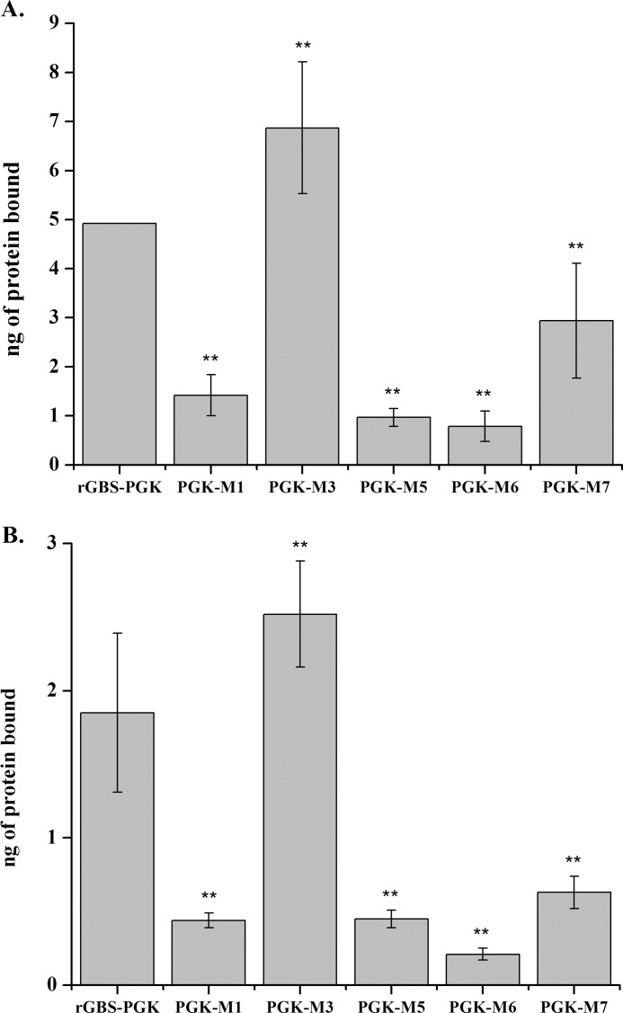
Phosphoglycerate kinase (PGK), present on the surface of group B streptococcus (GBS), has previously been demonstrated to bind the host proteins actin and plasminogen. The actin and plasminogen binding sites of GBS-PGK were identified using truncated GBS-PGK molecules, followed by peptide mapping. These experiments identified two actin and plasminogen binding sites located between amino acids 126-134 and 204-208 of the 398-amino acid-long GBS-PGK molecule. Substitution of the lysine residues within these regions with alanine resulted in significantly reduced binding to both actin and plasminogen. In addition, conversion of the glutamic acid residue at amino acid 133 to proline, the amino acid found at this position for the PGK protein of Streptococcus pneumoniae, also resulted in significantly reduced binding to actin and plasminogen. These results demonstrate that the lysine residues at amino acid positions 126, 127, 130, 204, and 208 along with the glutamic acid residue at amino acid position 133 are necessary for actin and plasminogen binding by GBS-PGK.


Figures

FIGURE 1.
FIGURE 1.
Binding of rGBS-PGK and TJB 1 to actin and plasminogen. Actin (1 μg/well) and plasminogen (0.1 μg/well) were fixed to wells of a 96-well polystyrene plate followed by incubation with 15 μg/ml rGBS-PGK or TJB 1 for 1 h. Wells were probed with anti-rGBS-PGK followed by anti-rabbit IgG alkaline phosphatase conjugate. A405 measurements were compared with standard curves to determine the amount of rGBS-PGK or TJB 1 remaining in the wells (supplemental Fig. S1). Data points represent the average value of experiments performed a total of nine times; error bars represent S.D.
FIGURE 2.
FIGURE 2.
Binding of plasminogen and actin to peptides generated based on the amino acid sequence of GBS-PGK. Peptides were resuspended in TBS and 15 μg/spot were fixed to a nitrocellulose membrane. A, membranes were incubated with actin (20 μg/ml) and probed with mouse anti-actin antibodies followed by goat anti-mouse IgG alkaline phosphatase conjugate antibodies; or, B, membranes were incubated with plasminogen (20 μg/ml) and probed with mouse anti-plasminogen antibodies followed by goat anti-mouse alkaline phosphatase conjugate antibodies. Images shown are representative of the experiments performed in triplicate. Spot C corresponds to rGBS-PGK (15 μg).
FIGURE 3.
FIGURE 3.
Binding of wild type and mutant GBS-PGK molecules to plasminogen and actin fixed to wells of a 96-well plate. Plasminogen (0.1 μg/well; A) or actin (1.0 μg/well; B) were fixed to wells of a 96-well plate followed by incubation with rGBS-PGK or rGBS-PGK containing mutations within the identified binding regions (15 μg/ml in blocking buffer). The amount of PGK remaining in the wells was assayed using rabbit anti-rGBS-PGK antibodies followed by goat anti-rabbit IgG alkaline phosphatase conjugate antibodies. The A405 readings were compared with standard curves containing 0–10 ng rGBS-PGK proteins to determine the amount of rGBS-PGK protein remaining in each well (supplemental Fig. S3). Data points represent the average value of experiments performed a total of nine times; error bars represent one S.D. **, statistical significance, p value < 0.01.
FIGURE 4.
FIGURE 4.
Actin binding to wildtype and mutated rGBS-PGK. rGBS-PGK and mutant rGBS-PGK were fixed to a 96-well polystyrene plate and then incubated with actin (20 μg/ml). Plates were probed with anti-actin antibodies followed by anti-mouse IgG alkaline phosphatase conjugate antibodies. Each data point represents the average value of experiments performed a total of six times; error bars represent one S.D. **, p value < 0.01 compared with the same concentration of non-mutated rGBS-PGK. The amount of each mutated rGBS-PGK required to bind the same amount of actin as 0.125 and 0.25 μg of non-mutated rGBS-PGK was determined to estimate the relative loss in binding activity. Lines indicate level of rGBS-PGK binding for comparison with mutants.
FIGURE 5.
FIGURE 5.
Plasminogen binding to wild type and mutated rGBS-PGK. rGBS-PGK and mutant rGBS-PGK were fixed to a 96-well polystyrene plate and then incubated with plasminogen (20 μg/ml). Plates were probed with mouse anti-plasminogen antibodies followed by goat anti-mouse IgG alkaline phosphatase conjugate antibodies. Data points represent the average of experiments performed a total of six times; error bars represent one S.D. **, statistical significance, p value < 0.01, compared with the same concentration of non-mutated rGBS-PGK. The amount of each mutated rGBS-PGK molecule required to bind the same amount of plasminogen as 0.125 and 0.25 μg of non-mutated rGBS-PGK was determine to estimate the relative loss in binding activity. Lines indicate level of rGBS-PGK binding for comparison with mutants.
FIGURE 6.
FIGURE 6.
Mapping the actin and plasminogen binding sites onto the model GBS-PGK structure. The amino acid sequence of GBS-PGK was submitted to iTASSER to generate a model structure. The model structure of GBS-PGK was visualized using RasMol. The regions of GBS-PGK identified to affect binding to actin and plasminogen through the site-directed mutagenesis experiment are highlighted on the model GBS-PGK molecule. Lysine residues are highlighted in red; glutamic acid residues are highlighted in blue.

Similar articles

Cited by

References

    1. Rubens C. E., Smith S., Hulse M., Chi E. Y., van Belle G. (1992) Respiratory epithelial cell invasion by group B streptococci. Infect. Immun. 60, 5157–5163 - PMC - PubMed
    1. Schuchat A., Robinson K., Wenger J. D., Harrison L. H., Farley M., Reingold A. L., Lefkowitz L., Perkins B. A. (1997) Bacterial meningitis in the United States in 1995. Active surveillance team. N. Engl. J. Med. 337, 970–976 - PubMed
    1. Verani J. R., Schrag S. J. (2010) Group B streptococcal disease in infants: Progress in prevention and continued challenges. Clin. Perinatol. 37, 375–392 - PMC - PubMed
    1. Weston E. J., Pondo T., Lewis M. M., Martell-Cleary P., Morin C., Jewell B., Daily P., Apostol M., Petit S., Farley M., Lynfield R., Reingold A., Hansen N. I., Stoll B. J., Shane A. J., Zell E., Schrag S. J. (2011) The burden of invasive early-onset neonatal sepsis in the United States, 2005–2008. Pediatr. Infect. Dis. J. 30, 937–941 - PMC - PubMed
    1. Edwards M. S., Baker C. J. (2005) Group B streptococcal infections in elderly adults. Clin. Infect. Dis. 41, 839–847 - PubMed

Publication types

MeSH terms

LinkOut - more resources