. 2012 Jun;8(6):e1002756.
doi: 10.1371/journal.pgen.1002756. Epub 2012 Jun 28.

G protein activation without a GEF in the plant kingdom

Affiliations

G protein activation without a GEF in the plant kingdom

Daisuke Urano et al. PLoS Genet. 2012 Jun.

Abstract

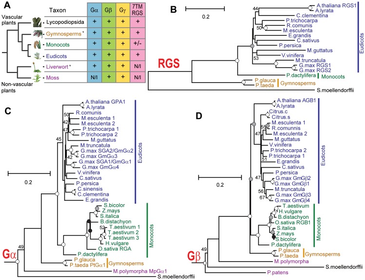
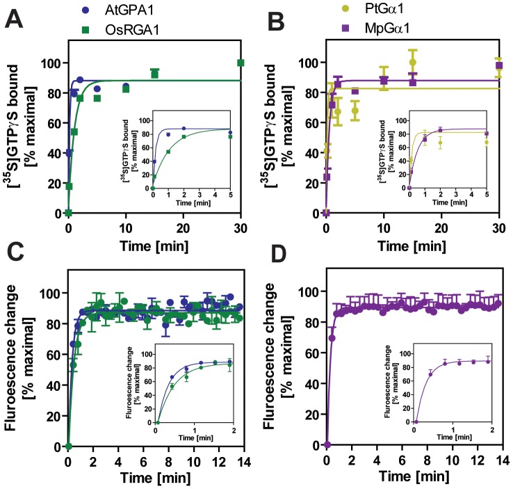
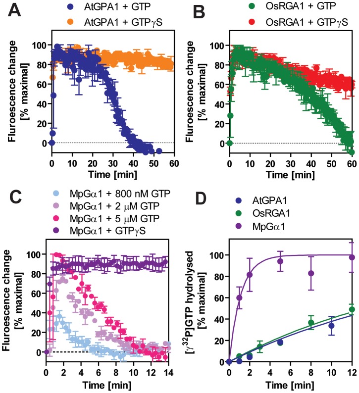
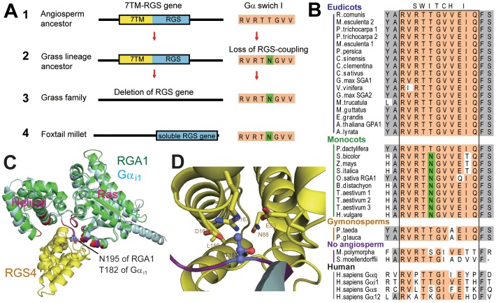
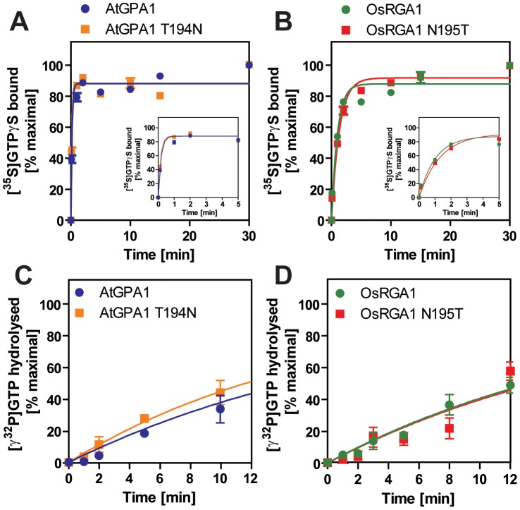
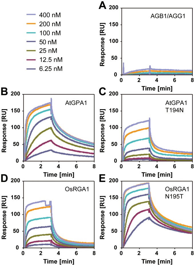
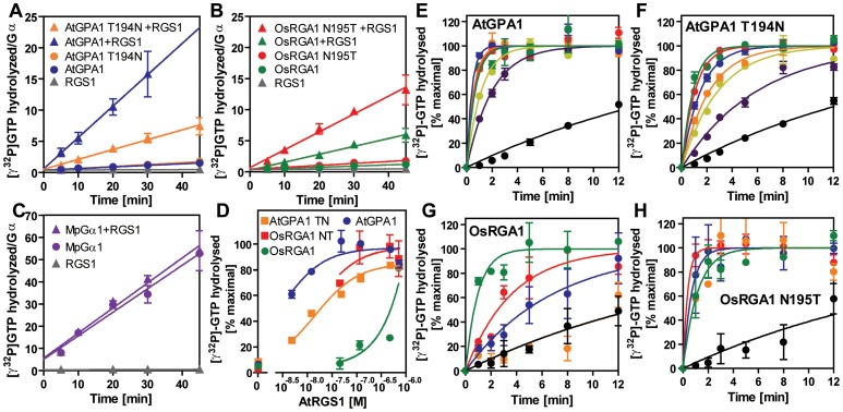
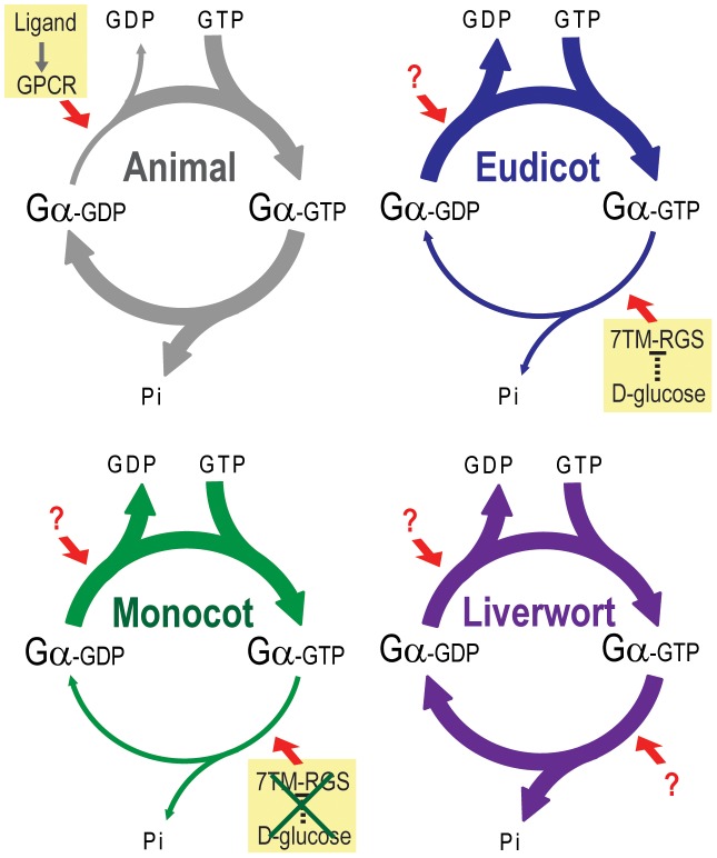
Animal heterotrimeric G proteins are activated by guanine nucleotide exchange factors (GEF), typically seven transmembrane receptors that trigger GDP release and subsequent GTP binding. In contrast, the Arabidopsis thaliana G protein (AtGPA1) rapidly activates itself without a GEF and is instead regulated by a seven transmembrane Regulator of G protein Signaling (7TM-RGS) protein that promotes GTP hydrolysis to reset the inactive (GDP-bound) state. It is not known if this unusual activation is a major and constraining part of the evolutionary history of G signaling in eukaryotes. In particular, it is not known if this is an ancestral form or if this mechanism is maintained, and therefore constrained, within the plant kingdom. To determine if this mode of signal regulation is conserved throughout the plant kingdom, we analyzed available plant genomes for G protein signaling components, and we purified individually the plant components encoded in an informative set of plant genomes in order to determine their activation properties in vitro. While the subunits of the heterotrimeric G protein complex are encoded in vascular plant genomes, the 7TM-RGS genes were lost in all investigated grasses. Despite the absence of a Gα-inactivating protein in grasses, all vascular plant Gα proteins examined rapidly released GDP without a receptor and slowly hydrolyzed GTP, indicating that these Gα are self-activating. We showed further that a single amino acid substitution found naturally in grass Gα proteins reduced the Gα-RGS interaction, and this amino acid substitution occurred before the loss of the RGS gene in the grass lineage. Like grasses, non-vascular plants also appear to lack RGS proteins. However, unlike grasses, one representative non-vascular plant Gα showed rapid GTP hydrolysis, likely compensating for the loss of the RGS gene. Our findings, the loss of a regulatory gene and the retention of the "self-activating" trait, indicate the existence of divergent Gα regulatory mechanisms in the plant kingdom. In the grasses, purifying selection on the regulatory gene was lost after the physical decoupling of the RGS protein and its cognate Gα partner. More broadly these findings show extreme divergence in Gα activation and regulation that played a critical role in the evolution of G protein signaling pathways.


Conflict of interest statement

The authors have declared that no competing interests exist.

Figures

Figure 1
Figure 1. Phylogeny of G protein components in the plant kingdom.
(A) Conservation of G protein components in the plant kingdom. *, Genomes have not been sequenced; N/I, Not identified; +/−, 7TM-RGS gene is identified in a date palm, but not in the grass-family. (B–D) Phylogram of the consensus maximum likelihood (ML) tree as determined for Gα, Gβ and multi transmembrane RGS protein sequences. The trees were rooted with Homo sapiensi1, Gαq, Gβ1, Gβ5 or S. moellendorffii 7TM-RGS genes. Bootstrap values above 40 are shown near each branch. White, gray or black circles indicate that the branch was supported more than 90%, 80% or 70%, respectively. Species and gene names are shown in blue, green, yellow or purple colors, indicating eudicots, monocots, gymnosperms or non-vascular plants, respectively. Scale bar represents 0.1 substitutions per sites. See Figure S1, S2, S3 for aligned sequences used for creating the trees.
Figure 2
Figure 2. Activation of plant Gα subunits.
(A, B) Time course of [35S]GTPγS binding to 1 µM Gα at 20°C. Data are presented as the mean ± SEM of more than triplicates. (C, D) Intrinsic tryptophan fluorescence of Gα was measured at room temperature. 5 µM GTPγS was added to 400 nM Gα in the cuvette at time 0. Data are mean ± SEM of duplicate samples.
Figure 3
Figure 3. Inactivation of plant Gα subunits.
(A–C) The activation and inactivation rates of Gα were monitored at room temperature by measuring the intrinsic tryptophan fluorescence. 800 nM GTP or 5 µM GTPγS was added to 400 nM Gα in the cuvette at time 0. Data are mean ± SEM of duplicate samples. Note that the turnover rate of Gα depends on stoichimometry of the concentrations of active Gα protein and GTP. The specific activity of the Gα subunits estimated by [35S]GTPγS binding assay were as follows: AtGPA1, 0.46 mol/mol protein; RGA1, 0.55 mol/mol protein; MpGPA1, 0.69 mol/mol protein. (D) Time course of single turnover [γ32P]GTP hydrolysis by 800 nM Gα. The mean ± SEM of duplicate samples is shown.
Figure 4
Figure 4. Evolution of monocot Gα and monocot specific loss of a 7TM-RGS gene.
(A) Evolutionary process to delete a 7TM-RGS gene in monocots. 1. The angiosperm ancestor possessed a 7TM-RGS gene. 2. After separation from the palm tree lineage, the Gα in the grass lineage lost a functional and physical coupling with its partner RGS protein. This uncoupling occurred by a single amino acid mutation, thus releasing the RGS gene from evolutionary constraint. 3. RGS genes were gradually deleted from grass-family genomes. 4. The extant S.italica RGS gene may still be functional, despite the loss of the 7TM region. (B) Switch I region of Gα. Conserved residues are highlighted with orange. Substituted Asn residue in monocots (Asn195 of OsRGA1) is shown with green. See Table S1 for species names. (C) Predicted structures of OsRGA1 (green) overlayed on Rattus norvegicusi1 and RGS4 (cyan and yellow, PDB: 1AGR [21]). Residues in purple (Gαi1) or red (OsRGA1) are at the binding surface to RGS4. Asn195 of OsRGA1 and Thr182 of Gαi1, discussed in this paper, are illustrated by a stick model. (D) The binding surface between Gαi1 and RGS4. Thr182 of Gαi1 and the interacting residues of RGS4 are shown in a stick model.
Figure 5
Figure 5. Intrinsic properties of Gα mutants.
(A, B) Time course of [35S]GTPγS binding to 1 µM Gα at 20°C. Data are presented as the mean ± SEM of triplicates. (C, D) Time course of single turnover [γ32P]GTP hydrolysis by 800 nM Gα. The mean ± SEM of duplicate samples is shown.
Figure 6
Figure 6. Affinity of plant Gα to AtRGS1 immobilized on the SPR biosensor.
Recombinant AtRGS1 (284–459 aa) was immobilized on sensor chip CM5. AlF4-bound Gα subunits or the Gβγ dimer control (A) flowed over the chip at seven different concentrations (6.25, 12.5, 25, 50, 100, 200, 400 nM). Kinetics determined with 1∶1 (Langmular) binding model is shown in Table 3. Wild type Arabidopsis Ga subunit (B), T194N mutant Arabidopsis Gα subunit (C), wild type rice Gα subunit (D), and 195T mutant rice Gα subunit (E).
Figure 7
Figure 7. GAP activity of RGS toward plant Gα subunits.
(A–C) Time course of steady-state [γ32P]GTP hydrolysis by 500 nM Gα in the presence or absence of 750 nM AtRGS1 were measured over time after incubation at 20°C. Rate of Pi production (mol/mol Gα protein) were shown. Data are mean ± SEM of duplicate samples. (D–H) Single turnover [γ32P]GTP hydrolysis by 500 nM Gα proteins with AtRGS1 (0 nM (black), 5 nM (purple), 12.5 nM (yellow), 50 nM (orange), 125 nM (blue), 500 nM (red) and 750 nM (green)). Data are mean +/− SEM for more than two individual experiments, except OsRGA1 with 50 nM RGS at 1 min and 5 min. Dose dependency of single time point (2 min) was shown in (D).
Figure 8
Figure 8. Model of G protein activation in the plant kingdom.
Slow rate of GDP release and GTP hydrolysis is indicated by a thin arrow. A rapid rate is indicated by a thick arrow. In animals, a rate of GDP release from Gα is much slower than that of GTP hydrolysis. Thus, acceleration of the GDP release by GPCR changes the G protein from inactive to active. In eudicots and monocots, GDP release is rapid, and GTP hydrolysis is much slower than the GDP release. Thus, G protein can self-activate without the aid of a GPCR or other GEF. Instead, the eudicot G protein is regulated by a 7TM-RGS protein, which constitutively promotes GTP hydrolysis step on plasma membrane. However, some monocot genomes lack the 7TM-RGS gene, thus some monocot G protein must use an unknown mechanism to regulate activation. In addition, a 7TM-RGS gene is not expressed in a liverwort. However, a liverwort G protein has a rapid rate of both GDP release and GTP hydrolysis, which is likely to compensate for the loss of the 7TM-RGS gene.

References

    1. Gilman AG. G proteins: transducers of receptor-generated signals. Annu Rev Biochem. 1987;56:615–649. - PubMed
    1. Sprang SR. G protein mechanisms: insights from structural analysis. Annu Rev Biochem. 1997;66:639–678. - PubMed
    1. Siderovski DP, Willard FS. The GAPs, GEFs, and GDIs of heterotrimeric G-protein alpha subunits. Int J Biol Sci. 2005;1:51–66. - PMC - PubMed
    1. Johnston CA, Taylor JP, Gao Y, Kimple AJ, Grigston JC, et al. GTPase acceleration as the rate-limiting step in Arabidopsis G protein-coupled sugar signaling. Proc Natl Acad Sci U S A. 2007;104:17317–17322. - PMC - PubMed
    1. Jones JC, Duffy JW, Machius M, Temple BR, Dohlman HG, et al. The crystal structure of a self-activating G protein alpha subunit reveals its distinct mechanism of signal initiation. Sci Signal. 2011;4:ra8. - PMC - PubMed

Publication types

MeSH terms