. 2012:2012:672013.
doi: 10.1155/2012/672013. Epub 2012 Jun 19.

Combined transfection of the three transcriptional factors, PDX-1, NeuroD1, and MafA, causes differentiation of bone marrow mesenchymal stem cells into insulin-producing cells

Affiliations

Combined transfection of the three transcriptional factors, PDX-1, NeuroD1, and MafA, causes differentiation of bone marrow mesenchymal stem cells into insulin-producing cells

Qing-Song Guo et al. Exp Diabetes Res. 2012.

Abstract

Aims: The goal of cell transcription for treatment of diabetes is to generate surrogate β-cells from an appropriate cell line. However, the induced replacement cells have showed less physiological function in producing insulin compared with normal β-cells.

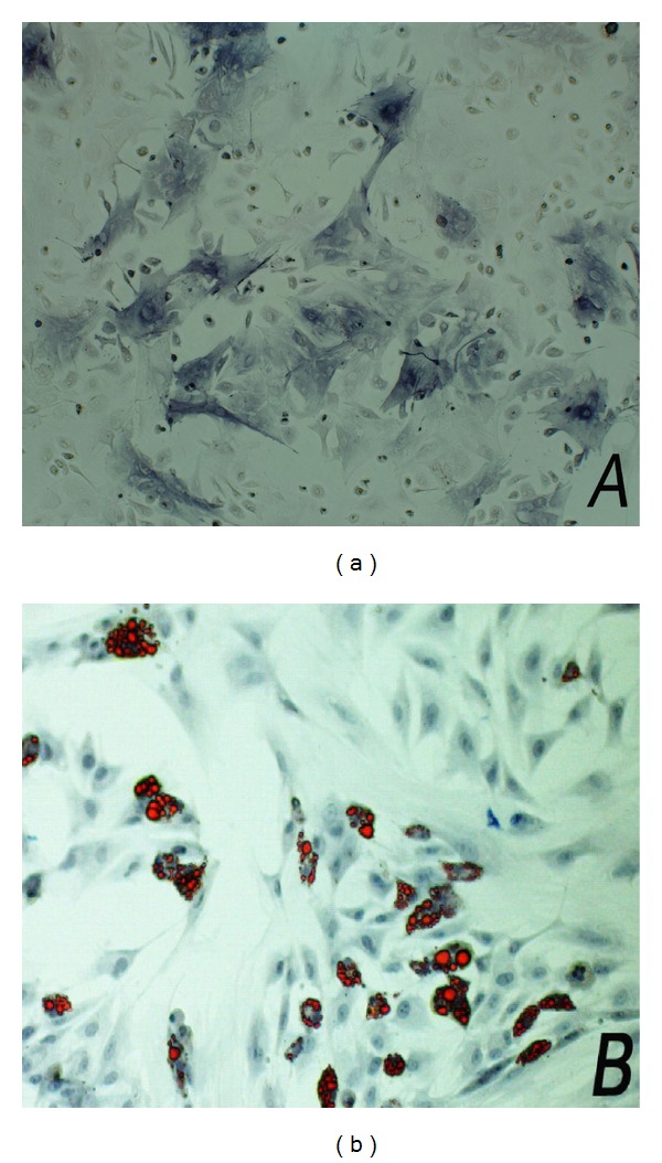
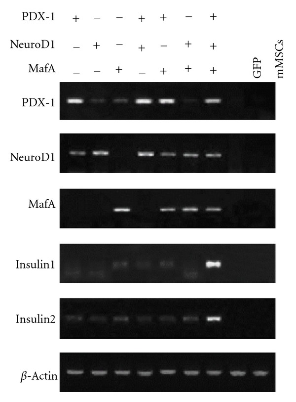
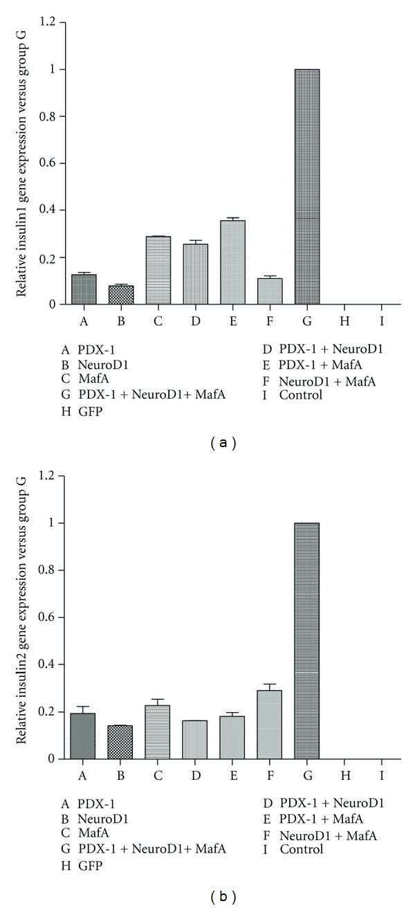
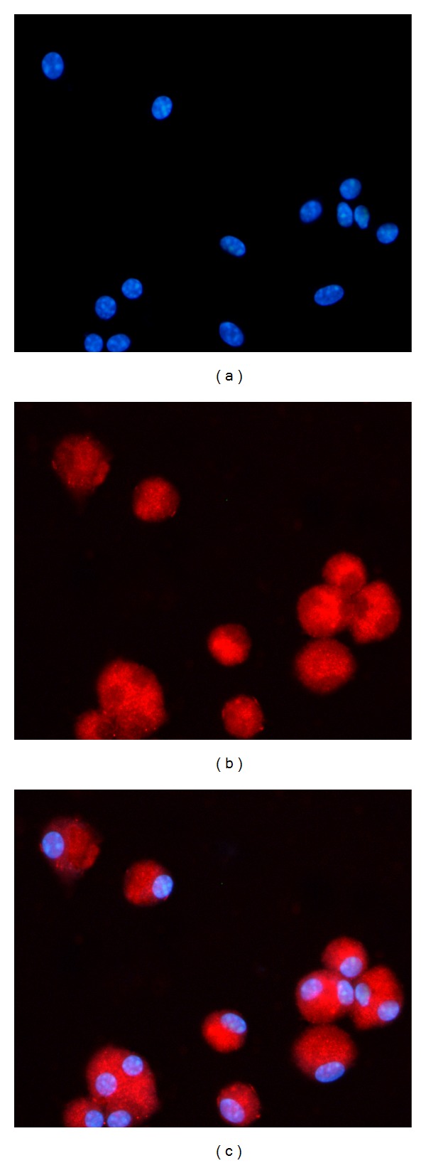
Methods: Here, we report a procedure for induction of insulin-producing cells (IPCs) from bone marrow murine mesenchymal stem cells (BM-mMSCs). These BM-mMSCs have the potential to differentiate into insulin-producing cells when a combination of PDX-1 (pancreatic and duodenal homeobox-1), NeuroD1 (neurogenic differentiation-1), and MafA (V-maf musculoaponeurotic fibrosarcoma oncogene homolog A) genes are transfected into them and expressed in these cells.

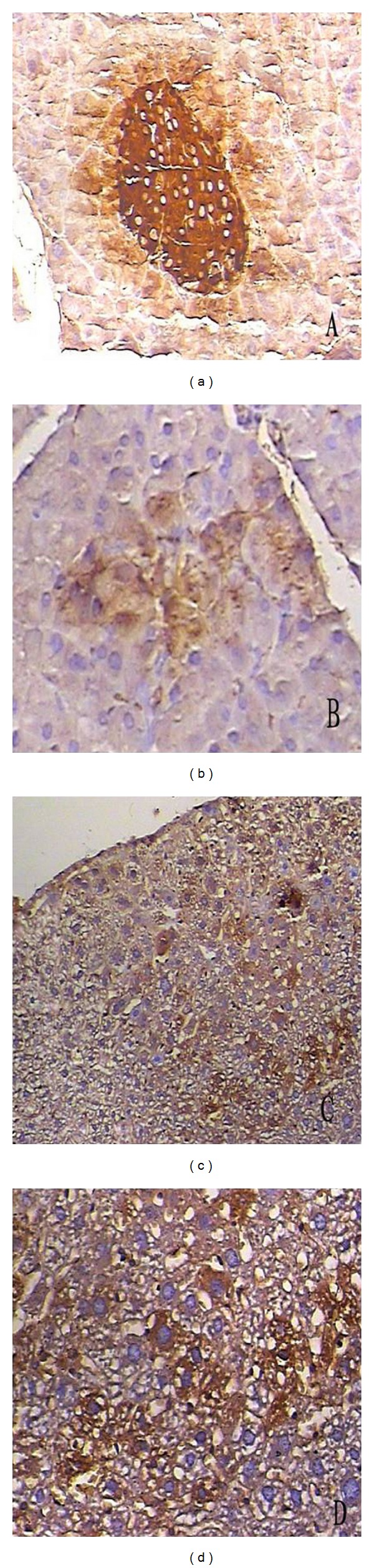
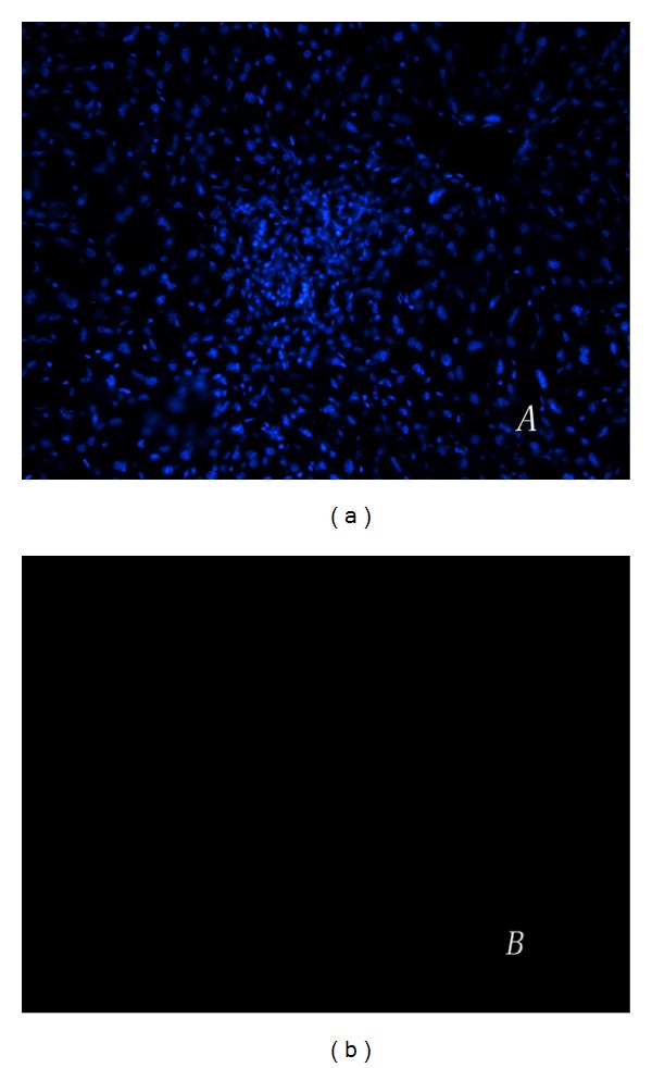
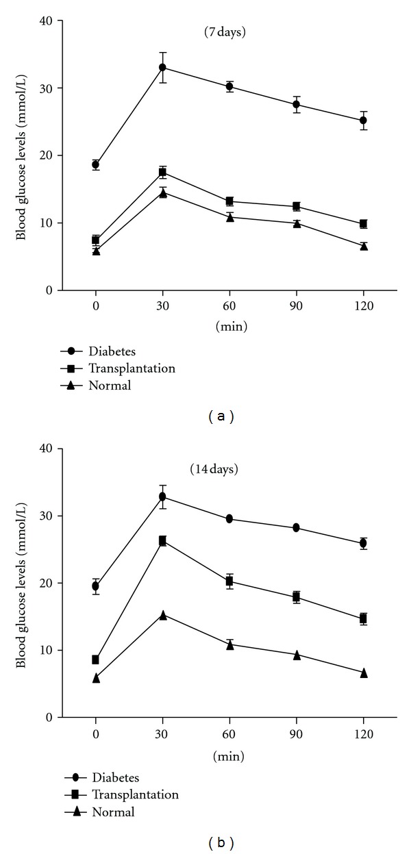
Results: Insulin biosynthesis and secretion were induced in mMSCs into which these three genes have been transfected and expressed. The amount of induced insulin in the mMSCs which have been transfected with the three genes together is significantly higher than in those mMSCs that were only transfected with one or two of these three genes. Transplantation of the transfected cells into mice with streptozotocin-induced diabetes results in insulin expression and the reversal of the glucose challenge.

Conclusions: These findings suggest major implications for cell replacement strategies in generation of surrogate β-cells for the treatment of diabetes.


Figures

Figure 1
Figure 1
mMSC were induced into osteoblasts and adipocytes in vitro under different differentiation medium. After incubation for 21 days, the differentiated cells were stained with alkaline phosphatase (a) or oil red (b) for their multipotent characteristic.
Figure 2
Figure 2
The toxic effect of adenovirus on mMSCs at different multiplicity of infection was determined by CCK-8. 10 μL CCK-8 solution was added into the same amount of mMSCs which were infected with Ad-GFP at different MOI. Optical density of each well varied directly with the survival of the cells. This experiment was repeated six times. Values are mean ± SD.
Figure 3
Figure 3
Adenovirus-mediated expression of PDX-1, NeuroD1, and MafA together induced expression of the insulin gene in infected mMSCs. mMSCs were infected with diverse single recombinant adenoviruses, both of the two adenoviruses, a combination of the three adenoviruses, or Ad-GFP. Total RNA from mMSCs was isolated 3 d after infection, and RT-PCR analysis was performed to examine expression of the specified genes. Cultured mMSCs without infection served as the negative control.
Figure 4
Figure 4
Quantitation of the amount of insulin1 and insulin2 that were produced by the infected mMSCs. The amount of insulin1 (a) and insulin2 (b) mRNA expression in mMSCs with triple infection was significantly larger compared with any other group.
Figure 5
Figure 5
Expression of insulin protein in the infected cells. After culturing them for 21 days, all the infected cells were incubated with anti-mouse insulin antibody. Nuclei were stained blue with Hoechst dye (a). Most of the transgenic cells were stained positively for insulin (b). In contrast, the mMSCs infected with Ad-GFP or null were negative for insulin.
Figure 6
Figure 6
Insulin secretion of the infected cells which were transferred into PDX-1, NeuroD1, MafA, or GFP at different differentiation stages in vitro. The cells were incubated in KRB containing the indicated concentration of glucose. The buffer was then collected for assay of insulin release in each experimental group. One asterisk, *P < 0.05. Data are presented as mean ± SD.
Figure 7
Figure 7
Immunohistochemistry assay for insulin of the survival infected mMSCs in the liver tissues of mice with diabetes. (a) Positive control, anti-mouse insulin staining of mouse pancreatic specimen showing an intense expression of insulin (enlargement ×100). (b) Anti-mouse insulin staining of pancreatic specimen of mice with STZ-induced diabetes showing a markedly decreased expression of insulin (enlargement ×100). (c) Infected mMSCs were injected into the livers of mice with diabetes three days after infection. The positive staining of mouse insulin expression can be clearly observed in the liver. (d) An enlargement of induced IPCs in the liver.
Figure 8
Figure 8
TUNEL assay was performed to see whether the injected cells were apoptotic. (a) Frozen tissue sections of the livers were stained with hoechst. (b) The immunofluorescent stainings of TUNEL were negative in the transplanted cells.
Figure 9
Figure 9
Glucose responses to glucose tolerance test of mice with diabetes after transplantation. The infected cells which expressed combination of PDX-1, NeuroD1, and MafA were transplanted into the livers of mice with STZ-induced diabetes, glucose tolerance test was performed at 7 (a) and 14 days (b) following transplantation, compared with a normal control and diabetes models without any treatment. Data are presented as mean ± SD.

Similar articles

Cited by

References

    1. Best M, Carroll M, Hanley NA, Piper Hanley K. Embryonic stem cells to β-cells by understanding pancreas development. Molecular and Cellular Endocrinology. 2008;288(1-2):86–94. - PubMed
    1. Kim JH, Shin KH, Li TZ, Suh H. Potential of nucleofected human MSCs for insulin secretion. Journal of Tissue Engineering and Regenerative Medicine. 2011;5(10):761–769. - PubMed
    1. Li G, Luo R, Zhang J, et al. Generating mESC-derived insulin-producing cell lines through an intermediate lineage-restricted progenitor line. Stem Cell Research. 2009;2(1):41–55. - PubMed
    1. Naujok O, Francini F, Picton S, Bailey CJ, Lenzen S, Jörns A. Changes in gene expression and morphology of mouse embryonic stem cells on differentiation into insulin-producing cells in vitro and in vivo. Diabetes/Metabolism Research and Reviews. 2009;25(5):464–476. - PubMed
    1. Bernardo AS, Hay CW, Docherty K. Pancreatic transcription factors and their role in the birth, life and survival of the pancreatic β cell. Molecular and Cellular Endocrinology. 2008;294(1-2):1–9. - PubMed

Publication types

MeSH terms

Substances