. 2012;7(6):e37251.
doi: 10.1371/journal.pone.0037251. Epub 2012 Jun 25.

Computational predictions of volatile anesthetic interactions with the microtubule cytoskeleton: implications for side effects of general anesthesia

Affiliations

Computational predictions of volatile anesthetic interactions with the microtubule cytoskeleton: implications for side effects of general anesthesia

Travis J A Craddock et al. PLoS One. 2012.

Abstract

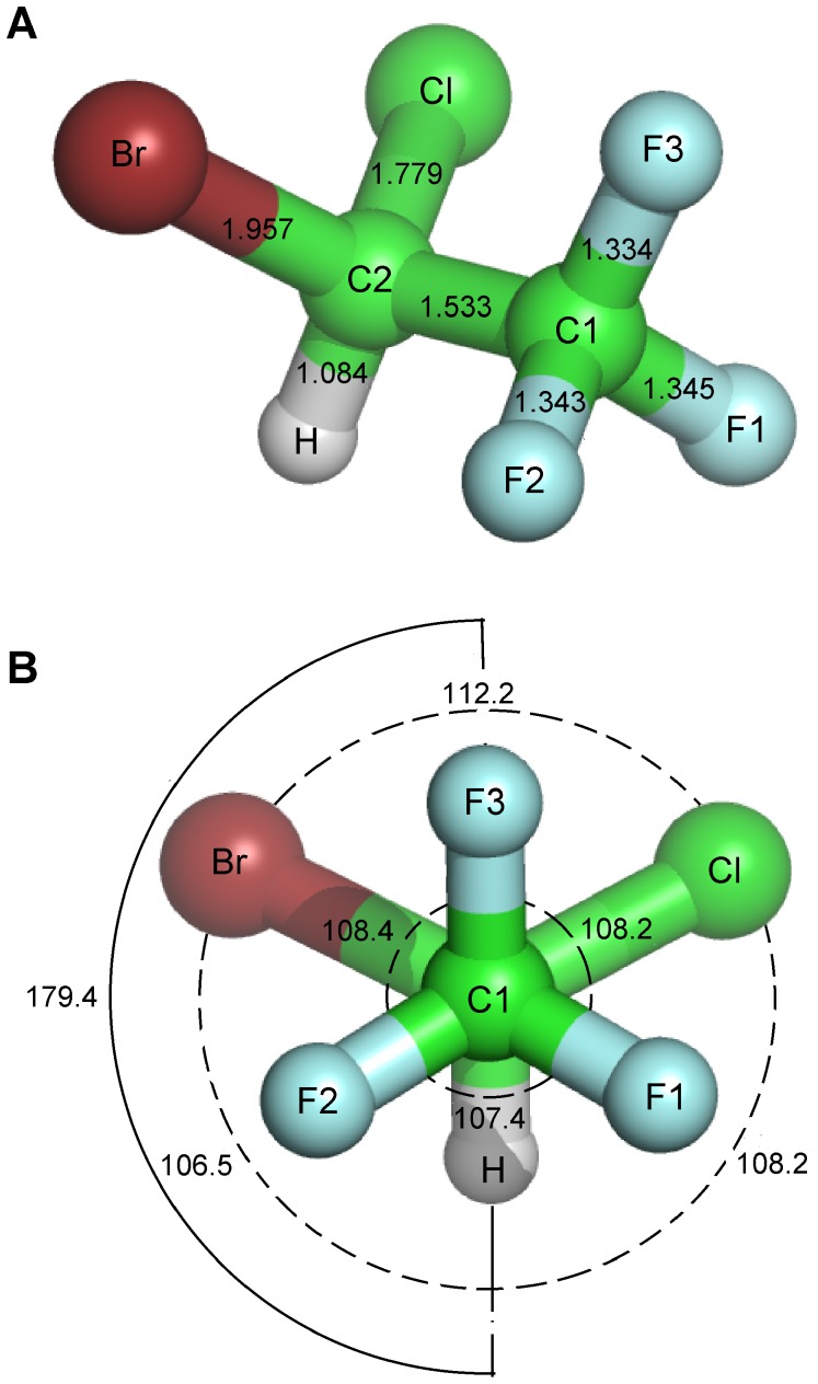
The cytoskeleton is essential to cell morphology, cargo trafficking, and cell division. As the neuronal cytoskeleton is extremely complex, it is no wonder that a startling number of neurodegenerative disorders (including but not limited to Alzheimer's disease, Parkinson's disease and Huntington's disease) share the common feature of a dysfunctional neuronal cytoskeleton. Recently, concern has been raised about a possible link between anesthesia, post-operative cognitive dysfunction, and the exacerbation of neurodegenerative disorders. Experimental investigations suggest that anesthetics bind to and affect cytoskeletal microtubules, and that anesthesia-related cognitive dysfunction involves microtubule instability, hyper-phosphorylation of the microtubule-associated protein tau, and tau separation from microtubules. However, exact mechanisms are yet to be identified. In this paper the interaction of anesthetics with the microtubule subunit protein tubulin is investigated using computer-modeling methods. Homology modeling, molecular dynamics simulations and surface geometry techniques were used to determine putative binding sites for volatile anesthetics on tubulin. This was followed by free energy based docking calculations for halothane (2-bromo-2-chloro-1,1,1-trifluoroethane) on the tubulin body, and C-terminal regions for specific tubulin isotypes. Locations of the putative binding sites, halothane binding energies and the relation to cytoskeleton function are reported in this paper.


Conflict of interest statement

Competing Interests: The authors have declared that no competing interests exist.

Figures

Figure 1
Figure 1. Tubulin in MT formation.
(A) Tubulin dimer. Light grey –α-tubulin, Dark Grey – β-tubulin. C-terminal tails extend from the main tubulin body. (B) B-lattice MT with protofilament highlighted. (C) Tubulin interactions in MT formation. Intradimer – between α- and β-tubulins, Longitudinal – between dimers in a protofilament, Lateral – between protofilaments.
Figure 2
Figure 2. Plot of protein backbone RMSD over 5 ns simulation.
Figure 3
Figure 3. Putative volatile anesthetic binding sites on the tubulin body.
(A) 47 total sites (red spheres) with persistence ranging from 0.80% to 100%. (B) 9 most persistent, and probable, sites (orange spheres), with persistence of 70% or greater.
Figure 4
Figure 4. Halothane molecule structure parameters.
(A) Bond lengths in Å. (B) Bond (dashed), and dihedral (solid) angles in degrees. Parameters obtained from an ab initio structure calculation .
Figure 5
Figure 5. Representative halothane binding modes on the TUBB2B C-terminal tail.
Red – N-terminal end connecting to the main tubulin body (body not shown for clarity), Blue – C-terminus. (A) −1.68 kcal/mol, (B) −2.3 kcal/mol, and (C) −2.79 kcal/mol.
Figure 6
Figure 6. Microtubule polymerization assays.
Black circle – General Tubulin Buffer (80 mM PIPES, MgCl2, 0.5 mM EGTA, pH 6.9). Green triangle – General Tubulin Buffer + 40 µM halothane. Blue square – General Tubulin Buffer + 10 µM paclitaxel. Red diamond – General Tubulin Buffer + 10 µM paclitaxel + 40 µM halothane. Mean values and standard deviation shown.
Figure 7
Figure 7. Halothane binding in the colchicine-binding pocket.
Blue – loop αT5, Red - strand βS9, Yellow – loop βT7 and helix βH8, Green – strand βS8, Orange – αE71, αN101, and βC241. (A) Colchicine binding site. (B) Halothane binding site 38, -2.72 kcal/mol, surrounded helix βH8 and strand βS8. (C) Halothane binding site 7, -3.13 kcal/mol, within 3 Å of βC241 and surrounded by strand βS9 and loop βT7.
Figure 8
Figure 8. Halothane binding in the vinblastine-binding pocket.
Red - loop αT7, Yellow – helix αH10, Green – strand αS9, Blue – loop βT5, Orange – helix βH6 and loop βH6-βH7. (A) Vinblastine binding site. (B) Halothane binding at site 21, −2.85 kcal/mol, within 2 Å of βY210 and surrounded by βH6 and βH7. (C) Halothane binding at site 39, −2.44 kcal/mol, within 5 Å of αK352 and βD179. (D) Halothane binding at site 1, −3.04 kcal/mol, within 3 Å of βD177 and surrounded by βT5.

References

    1. Campagna J, Miller K, Forman S. Mechanisms of actions of inhaled anesthetics. N Engl J Med. 2003;348:2110–2124. - PubMed
    1. Mihic SJ, Ye Q, Wick MJ, Koltchine VV, Krasowski MD, et al. Sites of alcohol and volatile anaesthetic action on GABAA and glycine receptors. Nature. 1997;389:385–389. - PubMed
    1. Franks NP, Lieb WR. Molecular and cellular mechanisms of general anaesthesia. Nature. 1994;367:607–614. - PubMed
    1. Franks NP, Lieb WR. Do general anaesthetics act by competitive binding to specific receptors? Nature. 1984;310:599–601. - PubMed
    1. Richards CD, Smaje JC. Anaesthetics depress the sensitivity of cortical neurones to L-glutamate. Br J Pharmacol. 1976;58:347–357. - PMC - PubMed

Publication types