. 2012;7(6):e38318.
doi: 10.1371/journal.pone.0038318. Epub 2012 Jun 25.

Crystal, solution and in silico structural studies of dihydrodipicolinate synthase from the common grapevine

Affiliations

Crystal, solution and in silico structural studies of dihydrodipicolinate synthase from the common grapevine

Sarah C Atkinson et al. PLoS One. 2012.

Abstract

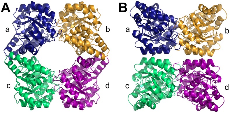
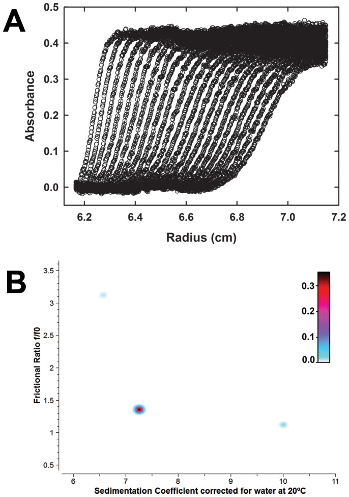
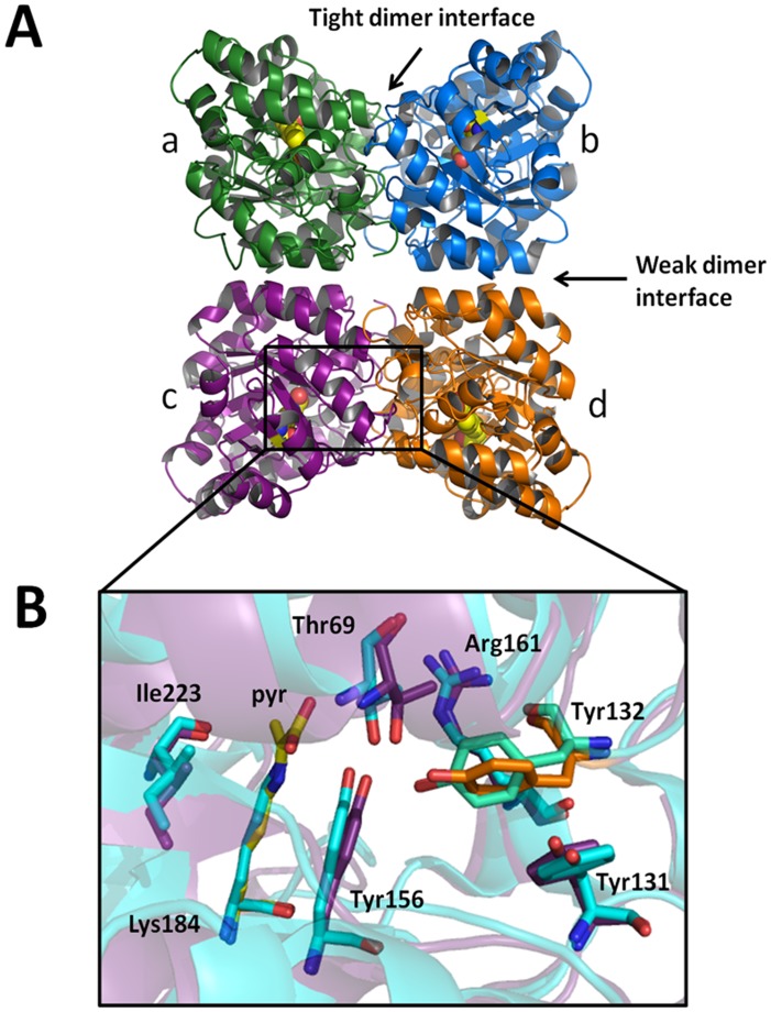
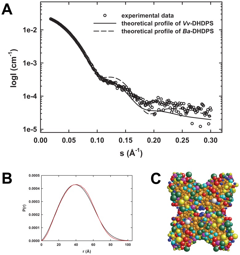
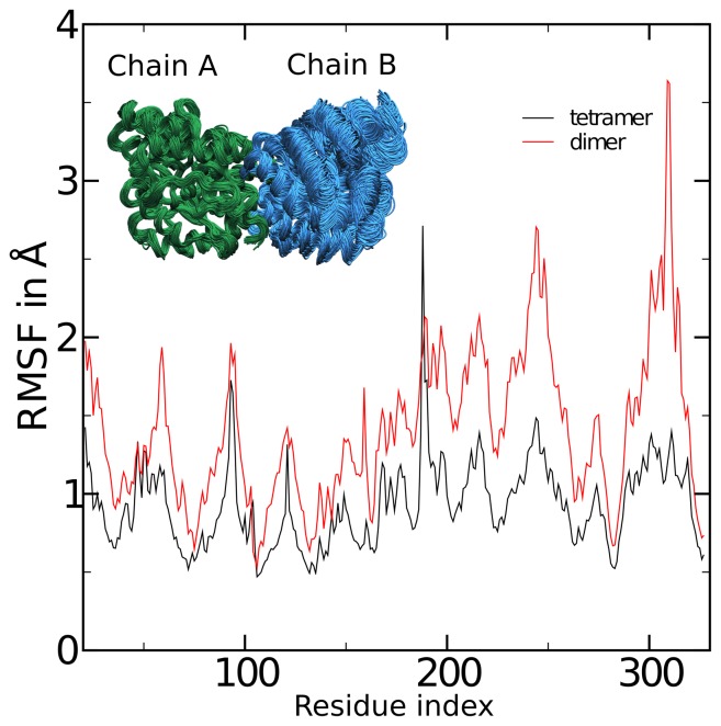
Dihydrodipicolinate synthase (DHDPS) catalyzes the rate limiting step in lysine biosynthesis in bacteria and plants. The structure of DHDPS has been determined from several bacterial species and shown in most cases to form a homotetramer or dimer of dimers. However, only one plant DHDPS structure has been determined to date from the wild tobacco species, Nicotiana sylvestris (Blickling et al. (1997) J. Mol. Biol. 274, 608-621). Whilst N. sylvestris DHDPS also forms a homotetramer, the plant enzyme adopts a 'back-to-back' dimer of dimers compared to the 'head-to-head' architecture observed for bacterial DHDPS tetramers. This raises the question of whether the alternative quaternary architecture observed for N. sylvestris DHDPS is common to all plant DHDPS enzymes. Here, we describe the structure of DHDPS from the grapevine plant, Vitis vinifera, and show using analytical ultracentrifugation, small-angle X-ray scattering and X-ray crystallography that V. vinifera DHDPS forms a 'back-to-back' homotetramer, consistent with N. sylvestris DHDPS. This study is the first to demonstrate using both crystal and solution state measurements that DHDPS from the grapevine plant adopts an alternative tetrameric architecture to the bacterial form, which is important for optimizing protein dynamics as suggested by molecular dynamics simulations reported in this study.


Conflict of interest statement

Competing Interests: Please note that co-authors Ashley M. Buckle, Juliet A. Gerrard, Renwick C.J. Dobson and Matthew A. Perugini are PLoS ONE Editorial Board members. This does not alter our adherence to all the PLoS ONE policies on sharing data and materials. Also note that co-authors Matthew T. Downton and John Wagner are employed by a commercial company - IBM Research Collaboratory for Life Sciences-Melbourne. This does not alter the authors' adherence to all the PLoS ONE policies on sharing data and materials.

Figures

Figure 1
Figure 1. DHDPS from bacteria and plants.
Dihydrodipicolinate synthase from (A) B. anthracis (PDB ID: 3HIJ [8]) and (B) N. sylvestris . Structural coordinates of the N. sylvestris DHDPS were kindly provided by Prof Robert Huber (Max Planck Institute for Biochemistry).
Figure 2
Figure 2. Sedimentation velocity analytical ultracentrifugation analysis of the quaternary structure of Vv-DHDPS in aqueous solution.
(A) Absorbance at 280 nm measured as a function of radial position from the axis of rotation (cm) for Vv-DHDPS (13 µM) centrifuged at 40,000 rpm. The raw data are presented as open symbols plotted at time intervals of 10 min overlaid with the 2DSA fit shown in panel B. (b) Pseudo-3D plots of solute distributions for 2DSA Monte Carlo of Vv-DHDPS using a grid resolution of 10,000 solutes. The colour scale represents the signal of each species in optical density units.
Figure 3
Figure 3. Crystal structure of Vv-DHDPS.
(A) Crystal structure of Vv-DHDPS (PDB ID: 3TUU) showing the position of the active site lysine residue (yellow spheres) in each monomer and the self-association interfaces. Two monomers come together at the tight dimer interface to form the dimeric unit, which dock at the weak dimer interface to form a homotetramer. The asymmetric unit contained eight monomers assembled as two homotetramers. (B) Active site residues of Vv-DHDPS overlaid with E. coli DHDPS (cyan). Pyruvate is shown in yellow. Tyr132 (orange) from the adjacent monomer interdigitates across the tight interface and is overlaid with the equivalent residue in E. coli DHDPS (green).
Figure 4
Figure 4. SAXS analyses of Vv-DHDPS.
(A) Theoretical scattering profiles from Vv-DHDPS (solid line) and Ba-DHDPS (dashed line) and the raw SAXS data (○).Theoretical scattering profiles were generated from crystallographic coordinates using CRYSOL. (B) P(r) plots of Vv-DHDPS from experimental data (black) and SOMO bead model (red) using ULTRASCAN. (C) SOMO bead model of Vv-DHDPS. The various colored beads represent acidic (green), hydrophobic (cyan), polar (red), basic (yellow) and non-polar (magenta) side-chains. Blue beads represent the protein main-chain and brown indicates buried beads.
Figure 5
Figure 5. Comparison of the molecular dynamics of the native tetramer and a putative dimeric form of Vv-DHDPS.
Simulations were analyzed by aligning chain A from all frames of the trajectories, and computing the root mean squared fluctuations (RMSF) of chain B, the monomer on the opposite side of the ‘tight-dimer’ interface. Shown are the RMSF values by residue number for the dimer (red) and tetramer (black). The inset shows 75 frames of the aligned dimer at 1 ns intervals.

References

    1. Dogovski C, Atkinson SC, Dommaraju SR, Hor L, Hutton CA, et al. Doelle H, editor. Lysine biosynthesis in bacteria – an unchartered pathway for novel antibiotic design. 2009. editor. Encyclopedia of life support systems, Volume 11 (Biotechnology Part I). Oxford: EOLSS Publishers. pp 116–136.
    1. Hutton CA, Perugini MA, Gerrard JA. Inhibition of lysine biosynthesis: an evolving antibiotic strategy. Mol Biosyst. 2007;3:458–65. - PubMed
    1. Dogovski C, Atkinson SC, Dommaraju SR, Downton M, Hor L, et al. Ekinci D, editor. Enzymology of bacterial lysine biosynthesis. Biochemistry: InTech Open Access Publisher. 2012. editor. (in press).
    1. Blickling S, Renner C, Laber B, Pohlenz HD, Holak TA, et al. Reaction mechanism of Escherichia coli dihydrodipicolinate synthase investigated by X-ray crystallography and NMR spectroscopy. Biochemistry. 1997;36:24–33. - PubMed
    1. Griffin MD, Dobson RC, Pearce FG, Antonio L, Whitten AE, et al. Evolution of quaternary structure in a homotetrameric enzyme. J Mol Biol. 2008;380:691–703. - PubMed

Publication types