. 2012;7(6):e39034.
doi: 10.1371/journal.pone.0039034. Epub 2012 Jun 27.

The safe use of a PTEN inhibitor for the activation of dormant mouse primordial follicles and generation of fertilizable eggs

Affiliations

The safe use of a PTEN inhibitor for the activation of dormant mouse primordial follicles and generation of fertilizable eggs

Deepak Adhikari et al. PLoS One. 2012.

Abstract

Background: Primordial ovarian follicles, which are often present in the ovaries of premature ovarian failure (POF) patients or are cryopreserved from the ovaries of young cancer patients who are undergoing gonadotoxic anticancer therapies, cannot be used to generate mature oocytes for in vitro fertilization (IVF). There has been very little success in triggering growth of primordial follicles to obtain fertilizable oocytes due to the poor understanding of the biology of primordial follicle activation.

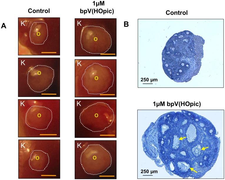
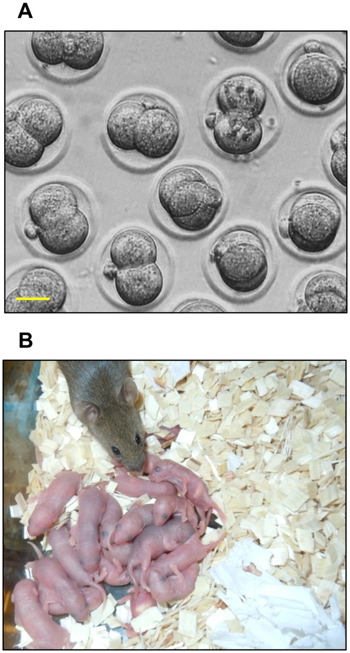
Methodology/principal findings: We have recently reported that PTEN (phosphatase and tensin homolog deleted on chromosome ten) prevents primordial follicle activation in mice, and deletion of Pten from the oocytes of primordial follicles leads to follicular activation. Consequently, the PTEN inhibitor has been successfully used in vitro to activate primordial follicles in both mouse and human ovaries. These results suggest that PTEN inhibitors could be used in ovarian culture medium to trigger the activation of primordial follicle. To study the safety and efficacy of the use of such inhibitors, we activated primordial follicles from neonatal mouse ovaries by transient treatment with a PTEN inhibitor bpV(HOpic). These ovaries were then transplanted under the kidney capsules of recipient mice to generate mature oocytes. The mature oocytes were fertilized in vitro and progeny mice were obtained after embryo transfer.

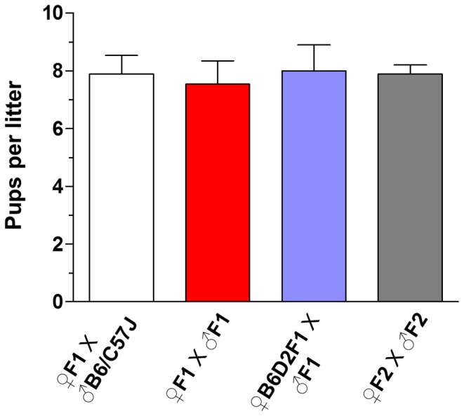
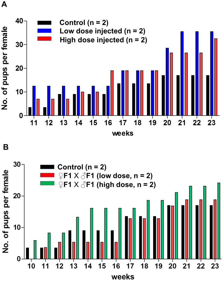
Results and conclusions: Long-term monitoring up to the second generation of progeny mice showed that the mice were reproductively active and were free from any overt signs or symptoms of chronic illnesses. Our results indicate that the use of PTEN inhibitors could be a safe and effective way of generating mature human oocytes for use in novel IVF techniques.


Conflict of interest statement

Competing Interests: The authors have declared that no competing interests exist.

Figures

Figure 1
Figure 1. Enhanced follicular development by transient treatment of neonatal mouse ovaries with the PTEN inhibitor bpV(HOpic).
(A) Comparison between the sizes of treated and control ovaries transplanted under the kidney capsules. One ovary from a PD3 mouse was cultured for 24 h with 1 µM bpV(HOpic) and another ovary was cultured without bpV(HOpic) and then transplanted under the capsule of each kidney of the same ovariectomized recipient as described in Materials and Methods. Ovaries that were treated with bpV(HOpic) before transplantation grew bigger than the non-treated control ovaries. K represents kidney tissue from the recipient, O represents the transplanted ovary, and the ovarian border is outlined by dashed circles. Scale bar = 1 mm. (B) Morphological analysis of treated and control ovaries excised from the kidney capsules. Ovaries from PD3 mice were cultured for 24 h with or without 1 µM bpV(HOpic) before transplantation under each kidney capsule of the same ovariectomized recipient as described in Materials and Methods. One day after the transplantation, recipient mice were treated daily with 2 IU of pregnant mare serum gonadotropin for 18 days. Fourteen hours before being killed, the mice were treated with 5 IU of human chorionic gonadotropin. Ovaries were excised from the kidney capsules and embedded in paraffin, and serial sections of 8 µm thickness were prepared and stained with hematoxylin. A larger number of antral follicles were observed in the bpV(HOpic)-treated ovaries (arrows) than in the control ovaries. The experiments were repeated at least 4 times, and 5 mice were used each time. Scale bar = 250 µm.
Figure 2
Figure 2. In vitro fertilization and birth of live pups after embryo transfer into the recipient mice.
(A) Fertilization of mature oocytes obtained from the PTEN inhibitor-treated ovaries. Ovaries from PD3 mice were cultured for 24 h with 1 µM bpV(HOpic) before transplantation under the kidney capsules of ovariectomized recipient mice as described in the Materials and Methods. One day after the transplantation, recipients were treated daily with 2 IU of pregnant mare serum gonadotropin for 18 days. After 18 days, the recipient mice were injected with 5IU human chorionic gonadotropin and the grafted ovaries were collected 14 h later. MII stage oocytes were fertilized in vitro as described in the Materials and Methods. After 24 h post fertilization, embryos had reached the two-cell stage. Scale bar = 25 µM. (B) Birth of live pups after embryo transfer. In vitro fertilized two-cell embryos were transferred into the oviducts of pseudopregnant surrogate mothers that had been prepared as described in the Materials and Methods.
Figure 3
Figure 3. Fertility measurement of the first and second generation progeny mice.
The F1 female and F1 male mice that were obtained by embryonic transfer were bred with B6/C57J male and B6D2F1 female mice, respectively. Fertility was also checked by breeding F1 males and F1 females. During the testing period, the mice regularly produced normal-sized F2 generation litters at normal intervals. To determine the fertility of the second generation mice, F2 females were bred with F2 males. n =  number of breeding pairs used.
Figure 4
Figure 4. Fertility measurement of the female mice directly injected with bpV(HOpic).
(A) Weekly comparison of the cumulative number of pups for the low dose bpV(HOpic)-injected mice (n = 2, blue bars), high dose-injected mice (n = 2, red bars) and control mice (n = 2, black bars). All mice had been bred with CD-1 strain males. (B) Weekly comparison of the cumulative number of pups produced by breeding the F1 mice. Breeding between F1 males and F1 females produced by low dose-injected females (n = 2, red bars), breeding between F1 males and F1 females produced by high dose-injected females (n = 2, green bars) and breeding between F1 males and F1 females produced by PBS-injected females (n = 2, black bars). n = number of breeding pairs used.

Similar articles

Cited by

References

    1. Hirshfield AN. Development of follicles in the mammalian ovary. Int Rev Cytol. 1991;124:43–101. - PubMed
    1. McGee EA, Hsueh AJ. Initial and cyclic recruitment of ovarian follicles. Endocr Rev. 2000;21:200–214. - PubMed
    1. Adhikari D, Liu K. Molecular mechanisms underlying the activation of mammalian primordial follicles. Endocr Rev. 2009;30:438–464. - PubMed
    1. Reddy P, Zheng W, Liu K. Mechanisms maintaining the dormancy and survival of mammalian primordial follicles. Trends Endocrinol Metab. 2010;21:96–103. - PubMed
    1. Beck-Peccoz P, Persani L. Premature ovarian failure. OrphanetJRareDis. 2006;1:9. - PMC - PubMed

MeSH terms