. 2012;7(6):e39174.
doi: 10.1371/journal.pone.0039174. Epub 2012 Jun 25.

Thaliporphine preserves cardiac function of endotoxemic rabbits by both directly and indirectly attenuating NFκB signaling pathway

Affiliations

Thaliporphine preserves cardiac function of endotoxemic rabbits by both directly and indirectly attenuating NFκB signaling pathway

A S Lee et al. PLoS One. 2012.

Abstract

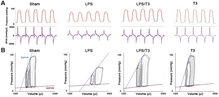
Cardiac depression in sepsis is associated with the increased morbidity and mortality. Although myofilaments damage, autonomic dysfunction, and apoptosis play roles in sepsis-induced myocardial dysfunction, the underlying mechanism is not clear. All of these possible factors are related to NFκB signaling, which plays the main role in sepsis signaling. Thaliporphine was determined to possess anti-inflammatory and cardioprotective activity by suppressing NFκB signaling in rodents. The purpose of this study is to further prove this protective effect in larger septic animals, and try to find the underlying mechanisms. The systolic and diastolic functions were evaluated in vivo by pressure-volume analysis at different preloads. Both preload-dependent and -independent hemodynamic parameters were performed. Inflammatory factors of whole blood and serum samples were analyzed. Several sepsis-related signaling pathways were also determined at protein level. Changes detected by conductance catheter showed Thaliporphine could recover impaired left ventricular systolic function after 4 hours LPS injection. It could also reverse the LPS induced steeper EDPVR and gentler ESPVR, thus improve Ees, Ea, and PRSW. Thaliporphine may exert this protective effect by decreasing TNFα and caspase3 dependent cell apoptosis, which was consistent with the decreased serum cTnI and LDH concentration. Thaliporphine could protect sepsis-associated myocardial dysfunction in both preload-dependent and -independent ways. It may exert these protective effects by both increase of "good"-PI3K/Akt/mTOR and decrease of "bad"-p38/NFκB pathways, which followed by diminishing TNFα and caspase3 dependent cell apoptosis.


Conflict of interest statement

Competing Interests: The authors have declared that no competing interests exist.

Figures

Figure 1
Figure 1. Thaliporphine could significantly ameliorate depressed hemodynamic characteristics of endotoxemic rabbits.
A. Pressure (top) and dP/dt (bottom) signals obtained with the pressure-volume conductance catheter system from sham, LPS (1 mg/kg), LPS plus 3 mg/kg Thaliporphine (LPS/T3), and 3 mg/kg Thaliporphine alone (T3) treated rabbits. B. Representative P-V loops at different preloads, showing differences in both end-systolic P-V relation (ESPVR) and end-diastolic P-V relation (EDPVR) between three groups. The less steep ESPVR and increased EDPVR in LPS treated animals indicate decreased contractile function and increased diastolic stiffness. Co-administration of 3 mg/kg Thaliporphine could reverse both cardiac depressive conditions.
Figure 2
Figure 2. Comparison of the protein expression of phospho-p38, total p38, phospho-p65, MMP9, and TNFα between sham, LPS, and LPS plus 3 mg/kg Thaliporphine (LPS/T3) groups.
Representative Western blots are shown at top A. and densitometric analyses at bottom B. The data of phospho-p65, MMP9, and TNFα were normalized with β–actin and phospho-p38 was normalized with total P38. Plots represent means+S.E. * P<0.05 as compared with sham group; # P<0.05 as compared with LPS group.
Figure 3
Figure 3. Comparison of the NFκB translocation between sham, LPS, LPS plus 3 mg/kg Thaliporphine (LPS/T3), and Thaliporphine alone (T3) groups.
A. Nuclear and cytosolic fractions were separately harvested for Western blot analysis of NFκB p65. B. The ratio of nuclear fraction of NFκB to cytosolic fraction was quantified. Plots represent means+S.E. * P<0.05 as compared with sham group; # P<0.05 as compared with LPS group. C. The translocation of NFκB p65 was performed with nuclear fractions in different groups by Electrophoretic mobility shift assay (EMSA). The arrows indicate the NFκB specific band and free probe. pc, positive control; ec, EMSA control (without probe); cp, cold (unlabeled) probe against the sample from LPS treated group.
Figure 4
Figure 4. Comparison of the protein expression of PI3K, phospho-Akt, total Akt, and mTOR between sham, LPS, and LPS plus 3 mg/kg Thaliporphine (LPS/T3) groups.
Representative Western blots are shown at top A. and densitometric analyses at bottom B. The data of PI3K and mTOR were normalized with β–actin and phospho-Akt was normalized with total Akt. Plots represent means+S.E. * P<0.05 as compared with sham group; # P<0.05 as compared with LPS group.
Figure 5
Figure 5. Thaliporphine significantly decreased cardiomyocytes apoptosis.
A. Apoptosis evidenced by TUNEL staining of apoptotic nuclei in different groups was compared. B. The morphology of cells was viewed under a light microscope. C. The fraction of apoptotic cells in heart tissue of all groups was quantified by 10 random fields in each slice (four slices in each group). D. Caspase3 activity was examined and expressed by means+S.E. * P<0.05 as compared with sham group; # P<0.05 as compared with LPS group.
Figure 6
Figure 6. Cytokine protein array analysis of serum from sham, LPS, LPS plus 3 mg/kg Thaliporphine (LPS/T3), and Thaliporphine alone (T3) groups.
Red circles indicate differences observed between sham and LPS. CINC-1, TIMP-1, and VEGF were markedly increased in LPS group but not both LPS/T3 and T3 group. The six signal points in the membrane of sham group indicate experimental positive controls.

Similar articles

Cited by

References

    1. Annane D, Bellissant E, Cavaillon JM. Septic shock. Lancet. 2005;365:63–78. - PubMed
    1. Martin GS, Mannino DM, Eaton S, Moss M. The epidemiology of sepsis in the United States from 1979 through 2000. N Engl J Med. 2003;348:1546–1554. - PubMed
    1. Hotchkiss RS, Karl IE. The pathophysiology and treatment of sepsis. N Engl J Med. 2003;348:138–150. - PubMed
    1. Poelaert J, Declerck C, Vogelaers D, Colardyn F, Visser CA. Left ventricular systolic and diastolic function in septic shock. Intensive Care Med. 1997;23:553–560. - PubMed
    1. Munt B, Jue J, Gin K, Fenwick J, Tweeddale M. Diastolic filling in human severe sepsis: an echocardiographic study. Crit Care Med. 1998;26:1829–1833. - PubMed

Publication types

MeSH terms