

Let-7b inhibits human cancer phenotype by targeting cytochrome P450 epoxygenase 2J2
- PMID: 22761738
- PMCID: PMC3382602
- DOI: 10.1371/journal.pone.0039197
Let-7b inhibits human cancer phenotype by targeting cytochrome P450 epoxygenase 2J2
Abstract
Background: MicroRNAs (miRNAs) are small, noncoding RNA molecules of 20 to 22 nucleotides that regulate gene expression by binding to their 3' untranslated region (3'UTR). Increasing data implicate altered miRNA participation in the progress of cancer. We previously reported that CYP2J2 epoxygenase promotes human cancer phenotypes. But whether and how CYP2J2 is regulated by miRNA is not understood.
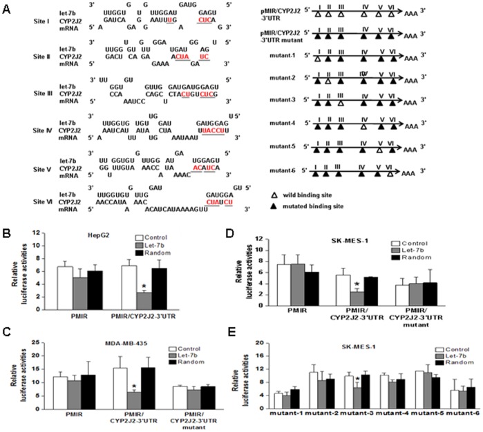
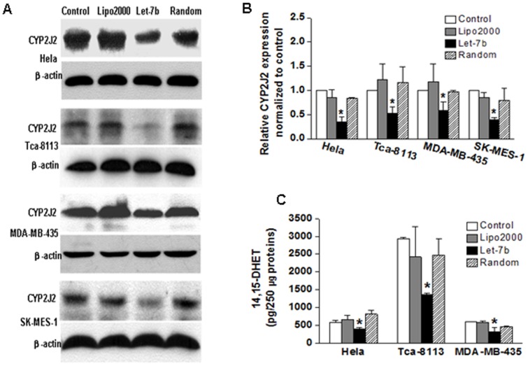
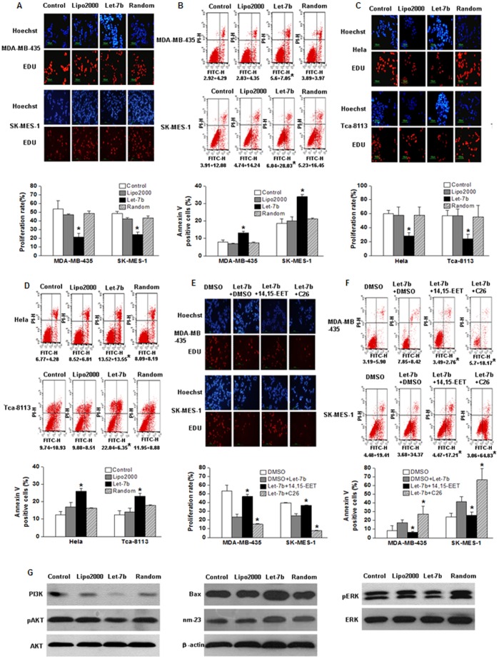
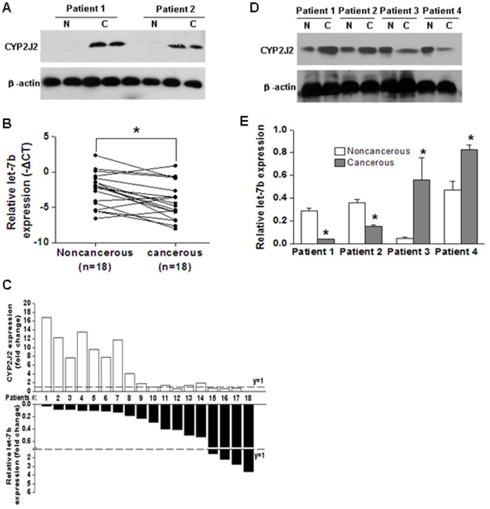
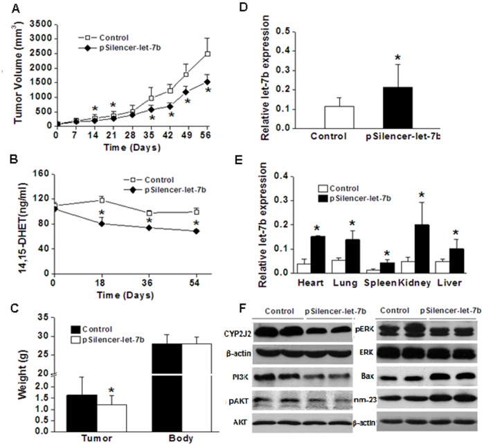
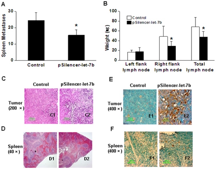
Methods and results: Using bioinformatics analysis, we found potential target sites for miRNA let-7b in 3'UTR of human CYP2J2. Luciferase and western blot assays revealed that CYP2J2 was regulated by let-7b. In addition, let-7b decreased the enzymatic activity of endogenous CYP2J2. Furthermore, let-7b may diminish cell proliferation and promote cell apoptosis of tumor cells via posttranscriptional repression of CYP2J2. Tumor xenografts were induced in nude mice by subcutaneous injection of MDA-MB-435 cells. The let-7b expression vector, pSilencer-let-7b, was injected through tail vein every 3 weeks. Let-7b significantly inhibited the tumor phenotype by targeting CYP2J2. Moreover, quantitative real-time polymerase chain reaction and western blotting were used to determine the expression levels of let-7b and CYP2J2 protein from 18 matched lung squamous cell cancer and adjacent normal lung tissues; the expression level of CYP2J2 was inversely proportional to that of let-7b.
Conclusions: Our results demonstrated that the decreased expression of let-7b could lead to the high expression of CYP2J2 protein in cancerous tissues. These findings suggest that miRNA let-7b reduces CYP2J2 expression, which may contribute to inhibiting tumor phenotypes.
Conflict of interest statement
Figures
Similar articles
-
Expression of cytochrome P450 arachidonic acid epoxygenase 2J2 in human tumor tissues and cell lines.Ai Zheng. 2009 Feb;28(2):93-6. Epub 2009 Feb 23. Ai Zheng. 2009. PMID: 19550113
-
let-7b and let-7c are determinants of intrinsic chemoresistance in renal cell carcinoma.World J Surg Oncol. 2015 May 8;13:175. doi: 10.1186/s12957-015-0596-4. World J Surg Oncol. 2015. PMID: 25951903 Free PMC article.
-
MicroRNA-30a suppresses breast tumor growth and metastasis by targeting metadherin.Oncogene. 2014 Jun 12;33(24):3119-28. doi: 10.1038/onc.2013.286. Epub 2013 Jul 15. Oncogene. 2014. PMID: 23851509
-
Cytochrome P450 2J2: distribution, function, regulation, genetic polymorphisms and clinical significance.Drug Metab Rev. 2013 Aug;45(3):311-52. doi: 10.3109/03602532.2013.806537. Drug Metab Rev. 2013. PMID: 23865864 Review.
-
[Research advances of artemin].Zhongguo Fei Ai Za Zhi. 2011 Oct;14(10):790-800. doi: 10.3779/j.issn.1009-3419.2011.10.05. Zhongguo Fei Ai Za Zhi. 2011. PMID: 22008109 Free PMC article. Review. Chinese. No abstract available.
Cited by
-
CYP2J2 overexpression increases EETs and protects against angiotensin II-induced abdominal aortic aneurysm in mice.J Lipid Res. 2013 May;54(5):1448-56. doi: 10.1194/jlr.M036533. Epub 2013 Feb 26. J Lipid Res. 2013. PMID: 23446230 Free PMC article.
-
The role and mechanisms of action of microRNAs in cancer drug resistance.Clin Epigenetics. 2019 Feb 11;11(1):25. doi: 10.1186/s13148-018-0587-8. Clin Epigenetics. 2019. PMID: 30744689 Free PMC article. Review.
-
Exosomes derived from microRNA-584 transfected mesenchymal stem cells: novel alternative therapeutic vehicles for cancer therapy.BMB Rep. 2018 Aug;51(8):406-411. doi: 10.5483/bmbrep.2018.51.8.105. BMB Rep. 2018. PMID: 29966581 Free PMC article.
-
Cardioprotective Effect of Decorin in Type 2 Diabetes.Front Endocrinol (Lausanne). 2020 Dec 7;11:479258. doi: 10.3389/fendo.2020.479258. eCollection 2020. Front Endocrinol (Lausanne). 2020. PMID: 33365011 Free PMC article.
-
Pharmacogenetics of Drug Metabolism: The Role of Gene Polymorphism in the Regulation of Doxorubicin Safety and Efficacy.Cancers (Basel). 2022 Nov 4;14(21):5436. doi: 10.3390/cancers14215436. Cancers (Basel). 2022. PMID: 36358854 Free PMC article. Review.
References
-
- Capdevila JH, Falck JR, Harris RC. Cytochrome P450 and arachidonic acid bioactivation: molecular and functional properties of the arachidonate monooxygenase. Journal of Lipid Research. 2000;41:163–181. - PubMed
-
- Wu S, Moomaw CR, Tomer KB, Falck JR, Zeldin DC. Molecular cloning and expression of CYP2J2, a human cytochrome P450 arachidonic acid epoxygenase highly expressed in heart. J Biol Chem. 1996;271:3460–3468. - PubMed
-
- Enayetallah AE, French RA, Thibodeau MS, Grant DF. Distribution of soluble epoxide hydrolase and of cytochrome P450 2C8, 2C9, and 2J2 in human tissues. J Histochem Cytochem. 2004;52:447–454. - PubMed
-
- Zeldin DC. Epoxygenase pathways of arachidonic acid metabolism. J Biol Chem. 2001;276:36059–36062. - PubMed
-
- Karara A, Makita K, Jacobson HR, Falck JR, Guengerich FP, et al. Molecular cloning, expression, and enzymatic characterization of the rat kidney cytochrome P-450 arachidonic acid epoxygenase. J Biol Chem. 1993;268:13565–13570. - PubMed
Publication types
MeSH terms
Substances
LinkOut - more resources
Full Text Sources
Medical
Miscellaneous
